|
Abstract
Bezoars are concretions of swallowed hair, fruit vegetable fibers, and similar substances found in the alimentary canal. The first description of a postmortem human bezoar was by Swain in 1854. Although the prevalence of bezoars in humans is low, an absence of treatment has been associated with mortality rates as high as 30%, primarily because of gastrointestinal bleeding, destruction, or perforation.
Introduction
This is a report describing acute intestinal obstruction due to a phytobezoar following truncal vagotomy and gastrojejunostomy in
a 68-year-old man.
Case Report
A 68 year-old male who had undergone a gastrojejunostomy seven years earlier as part of his surgical management for a duodenal ulcer refractory to medical treatment presented with epigastric discomfort, abdominal pain and vomiting of 10 days duration. Physical examination showed that he was afebrile, with a pulse rate of 138 beats/min and a respiratory rate of 28/min. Chest and cardiac findings were unremarkable. The abdomen was soft and non-tender and not distended. No focal neurologic deficits were apparent.
Laboratory test results showed a hemoglobin concentration of 16.9 g/dL, a leukocyte count of 13 x 109 /L, and a platelet count of 351 x 109 /L. His serum creatinine was 366 mg/dl, his urea nitrogen was 24.5 mg/dl, his Na concentration was 130 mEq/L and his serum glucose concentration was 25.11 mg/dl. Upper GI contrast study showed a persistently dilated proximal jejunal loop. (Fig. 1)
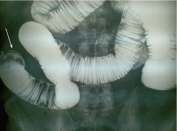
Figure 1: Barium study showing mid-jejunal opacity with a claw- like appearance. The bowel loop distal to the filling defect has collapsed (arrow).
Despite the administration of intravenous fluids and cessation of oral feeding, the patient continued to vomit continuously. An exploratory laparotomy through an upper midline abdominal incision revealed a hard, mobile mass causing a complete obstruction in the jejunum. The mass was exteriorized through an enterotomy, (Fig. 2). The mass had a rough, greenish-black outer surface, which appeared whitish due to the contrast used in the upper GI study. His postoperative period was unremarkable, other than mild vomiting during the first two days.
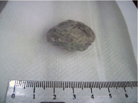
Figure 2: Removal of the bezoar through laparotomy and enterotomy from the mid-loop of the jejunu
Discussion
Bezoars occur most frequently in patients with a previous history of gastric operation and are detected in up to 20% of patients who have undergone antrectomy.1 Phytobezoar formation may be due to a reduction in gastric acidity, peptic activity, poor gastric mixing, and/or delayed emptying.2
The diagnosis of a gastric trichobezoar can be confirmed by radiography or endoscopy. Plain films of the abdomen may reveal amorphous, granular, calcified, or whirlpool-like configurations of solid and gaseous material within the stomach.3 Unlike phytobezoars, which are generally impervious to barium, trichobezoars tend to absorb barium, aiding in their diagnosis. The current gold standard for diagnosis of bezoars is upper gastrointestinal endoscopy, which provides direct visualization of the bezoar and allows sample taking and therapeutic intervention.4
Sonographic evidence of an intraluminal mass with a hyper- echoic arc-like surface and a marked acoustic shadow, suggestive of a bezoar, has been reported.5,6 Moreover, the marked acoustic shadowing behind the echogenic band produced by a bezoar differs from the "dirty" shadowing generated by ingested gas and food within the stomach.7 Since bezoars produce the same sonographic images as ectopic lithiasis, it is important to distinguish bezoar- induced obstructions from gallstone ileus.8
Bezoars have a characteristic appearance on CT, usually presenting as a well circumscribed in-homogeneous intraluminal mass with a mottled gas pattern in the dilated small bowel at the site of obstruction and abrupt collapse of the lumen beyond the lesion.9,10
Conclusion
Therapy for any bezoar necessitates its removal and prevention of recurrence. Small bezoars may be amenable to naso-gastric lavage or suction, a clear liquid diet, and the use of prokinetic agents.11 Larger bezoars may be fragmented mechanically or with digestive enzymes.12 Endoscopic retrieval and fragmentation are frequently used for proximal bezoars whose size and density are not prohibitive; however, the procedure can be technically challenging, and fragments may migrate distally and cause small bowel obstruction.13
A recently described technique from China incorporates a laser mini-explosive technique through an endoscope; in 100 patients, the cure rate was 100%.14 Laparotomy is reserved for bezoars that have caused perforation (7%) or hemorrhage (10%), or that are too large or obstructive to be managed less invasively.3 In patients who have undergone gastrectomy, however, the recurrence rate of phytobezoars is 13.5%, despite preventive measures.1
Acknowledgements
The author reported no conflict of interest and no funding was received on this work.
|