A 56-year-old woman presented with an incidentally detected calcification in the left kidney following ultrasonography, which was performed for the evaluation of right sided upper abdominal pain. Ultrasound also showed multiple gall bladder calculi. Their were no other abnormalities. The patient did not report any history of pain over the left flank, or any urinary complaints. The patient was treated for pulmonary tuberculosis at the age of 25. Routine urine microscopic examination and abdominal examination were normal, and there were no enlarged peripheral lymph nodes. Blood urea was 45mg/dL and serum creatinine was 0.9mg/dL. A kidney, ureter, and bladder (KUB) X-ray and computed tomography (CT) urography is shown in Figure 1 and Figure 2, respectively.
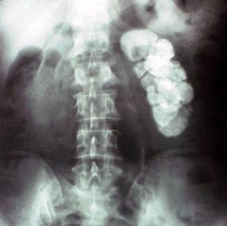
Figure 1: Kidney, ureter, and bladder (KUB) X-ray
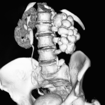
Figure 2: Computed tomography (CT) urography
Questions
- What is the diagnosis?
- What should the treatment be?
Answers
- The diagnosis is putty kidney with autonephrectomy secondary to tuberculosis. KUB x-ray (Fig.1) shows calcification of the left kidney. CT urography confirms that the entire substance of the left kidney is replaced by a dense calcification and there is no excretion of contrast from the left kidney (Fig.2).
- No treatment is required in asymptomatic patients; however, nephrectomy is indicated if there is pain, hypertension alongside a unilateral renal lesion, or recurrent urinary tract infections.
Discussion
A chest X-ray of the patient was normal, and centrifuged urine sample was negative for AFB (acid-fast bacilli). As the patient was not symptomatic and blood pressure was normal, nephrectomy was not considered. She underwent laparoscopic cholecystectomy for gallstones. Genitourinary tract is the most common site of extrapulmonary involvement of tuberculosis (TB) Renal TB usually occurs secondary to haematogenous dissemination of Mycobacterium tuberculosis from the lungs.1 Tubercle bacilli lodge in periglomerular capillaries and form cortical granulomas that can remain stable for many years.2 If reactivation occurs, the organisms spread into the medulla, causing papillary necrosis. As the disease progresses, extensive papillary necrosis may present with the formation of frank cavities destroying the renal parenchyma. Communication of the granulomas with the collecting system can lead to spreading of bacilli into the renal pelvis, ureter, urinary bladder, and genital organs. With complete obstruction due to tuberculous ureteric stricture, tuberculous pyonephrosis with parenchymal destruction and caseation may occur, which is calcified. This is called putty kidney, also known as chalked kidney or cement kidney, and results in autonephrectomy. Urine tests are negative for Mycobacterium tuberculosis, though there are mycobacteria in the destroyed kidney. Renal tuberculosis usually develops 10 to 20 years after the initial infection3 and may be confused with renal stones. Indication of nephrectomy includes pain, hypertension alongside a unilateral renal lesion, and recurrent urinary tract infections.4
references
- Becker JA. Renal tuberculosis. Urol Radiol 1988;10(1):25-30.
- Leder RA, Low VH. Tuberculosis of the abdomen. Radiol Clin North Am 1995 Jul;33(4):691-705.
- Kenney PJ. Imaging of chronic renal infections. AJR Am J Roentgenol 1990 Sep;155(3):485-494.
- Clifford AV, Noon AP, Raw D, Hall J. Renal calcified mass misdiagnosed as a renal calculus in an adult with tuberculosis “autonephrectomy”: a case report. Cases J 2009;2:7613.