|
Abstract
External jugular vein aneurysm with thrombosis presenting as neck swelling is a rare clinical entity and rarely encountered in routine clinical practice. We present a case of a 45-year-old female with external jugular vein aneurysm presenting as a painful lump in the neck. Ultrasound of the neck and CT angiography showed saccular dilation of the lower end of the external jugular vein with thrombosis within the aneurysm. Saccular aneurysm of the external jugular vein is very uncommon and can lead to thrombotic complications with serious consequence.
Keywords: Saccular aneurysm; External jugular vein; Thrombosis; Painful neck lump.
Introduction
Venous aneurysms of the neck are rare clinical entities. Venous aneurysm of the neck commonly involves the internal jugular vein.1 Venous aneurysm of external jugular vein is very rare and very few cases have been reported in the English literature.2-7 Aneurysmal dilatations of the cervical venous system are rare due to the low pressure in the vena cava.8 Common etiologic factors are tumor, thoracic outlet syndrome and trauma.6 Inflammation, degeneration, and increased pressure within the venous system could also be possible causes of venous aneurysm in the neck.5 Venous aneurysms in the neck usually have a benign clinical course and may present as cervical swelling, pain and tenderness in the neck.
In this report, we describe a 45-year-old female with aneurysm of the external jugular vein and its clinical and radiological characteristics.
Case Report
A 45-year-old woman presented at our outpatient department with complaints of progressive swelling on left supraclavicular region for 3 months. The swelling had increased in size during the last 15 days with accompanying pain and feeling of tightness over the left side of the neck. There was no history of preceding trauma, and there was no history of difficulty in swallowing, change in voice or any nasal symptoms. Furthermore, there was no history of prior hospital admission.
Physical examination revealed a 3 × 3 cm soft, round, nonpulsatile, fluctuant swelling on the left supraclavicular region. The overlying skin over the swelling had no signs of inflammation. Swelling was mobile in horizontal plane. There was mild increase in the size of the swelling during Valsalva manoeuvre and during laughing. The swelling was mildly compressible and slip sign was negative. No bruit could be detected on auscultation over the swelling. There was no other swelling in the neck or on other parts of the body. (Fig. 1)
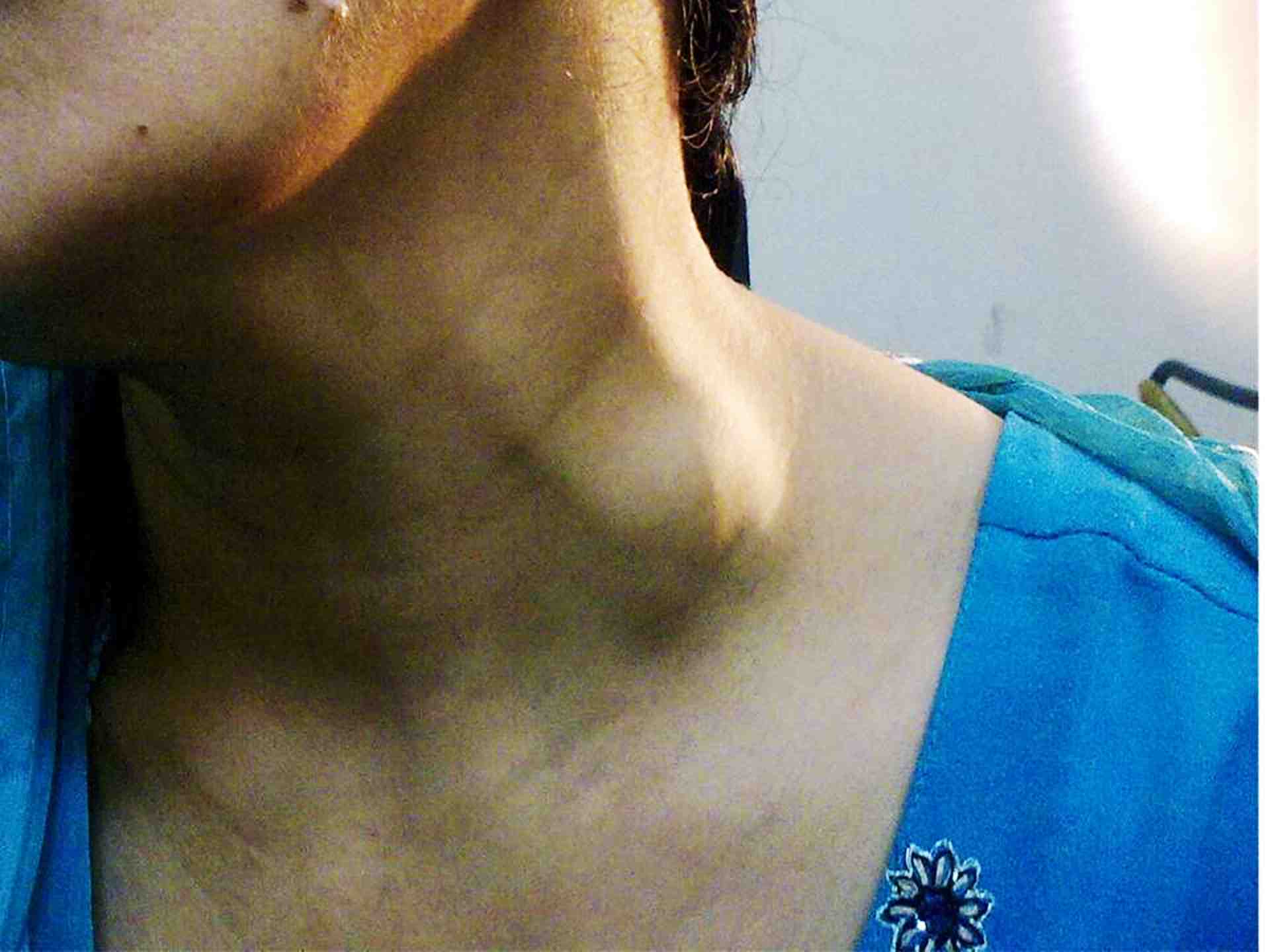
Figure 1: A 45-year-old female with lump in left supraclavicular region for three months.
Ultrasonography of the neck reported a thin-walled cystic mass on the left side abutting the left external jugular vein, (Fig. 2). Contrast-enhanced computed tomography scan of the neck showed a focal area of dilatation of the left external jugular vein and enhancing oval-shaped swelling arising from the external jugular vein with calcification at the periphery of the lesion. (Fig. 3)
The patient was reassured and asked to come for regular followup. She returned after a week with complaints of increasing pain over the swelling. A decision to excise the swelling was made. Under general anesthesia, adhesion to the surrounding tissues was freed and the mass was excised after ligating the proximal and distal ends of the external jugular vein, (Fig. 4). Postoperative period was uneventful.
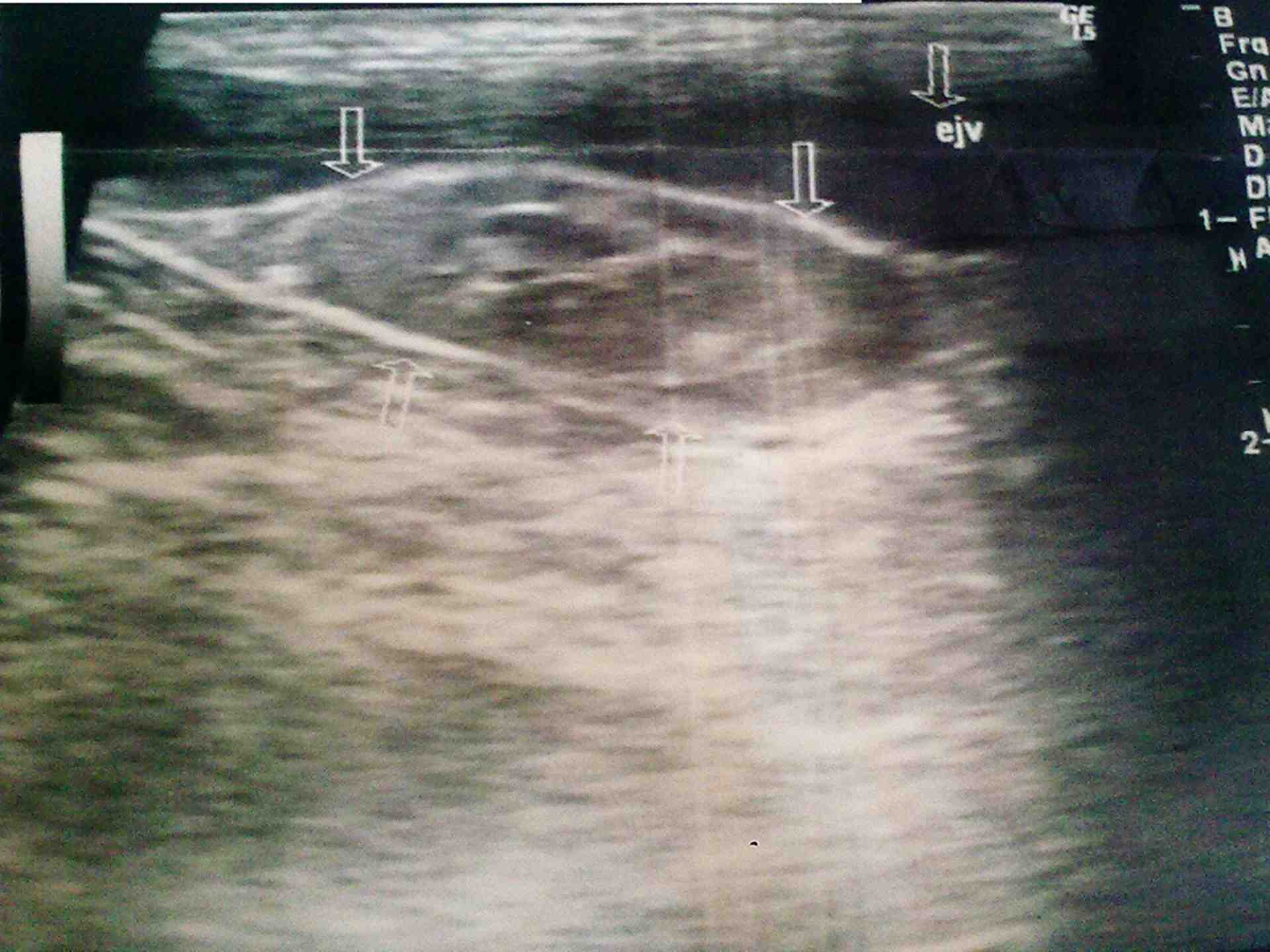
Figure 2: USG of neck showing aneurysmal dilation of external jugular vein.
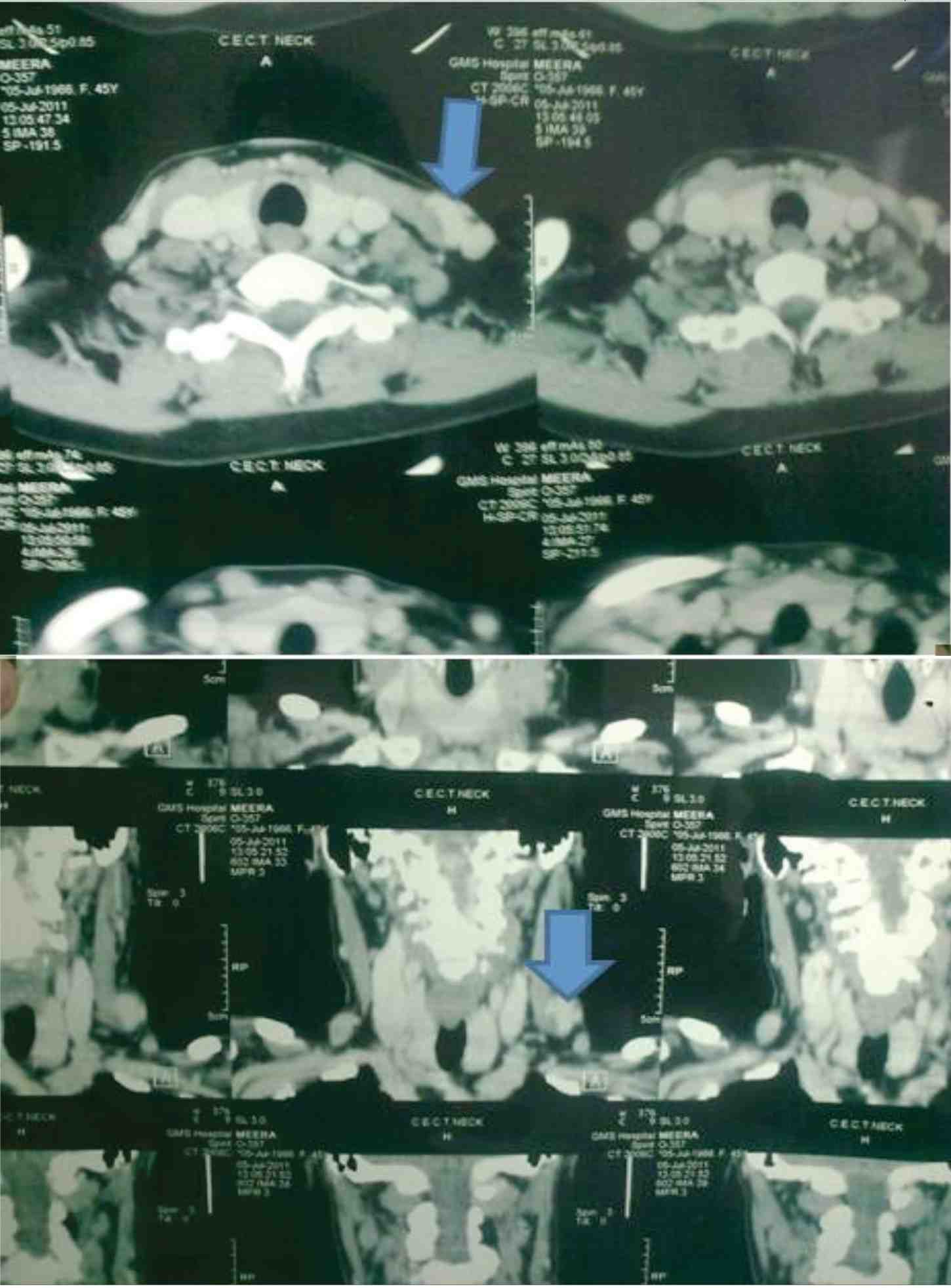
Figure 3: CT scan of the neck showing aneurysmal dilation of external jugular vein (arrow) with areas of calcification within the mass.
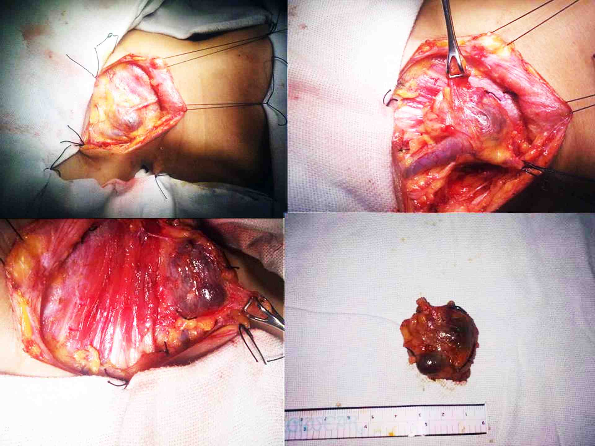
Figure 4: Resection of aneurysm with the excised mass.
Histopathology of the excised mass showed a grossly dilated vein, 3 × 2 × 1 cm globular shape, and the presence of a thrombus inside the dilated vein. Microscopically, it showed a dilated intact venous wall with marked reduction of smooth muscle cells in the media. A diagnosis of venous aneurysm was made.
Discussion
Venous aneurysm was first described in the literature by Harris in 1928.9 Hischler suggested the term venous aneurysm similar to arterial aneurysm.10 True venous aneurysms are rarely encountered compared to arterial aneurysm, as well as can affect any veins including intracranial, cervical, thoracic, visceral, and upper and lower extremity veins; however, venous aneurysms of the neck are rare due to low pressure in the vena cava system.7 In the neck, the internal jugular vein is the most common vein involved.1 Aneurysm of the external jugular vein is rare and very few cases have been reported in the literature to date.
Venous aneurysm can be congenital or acquired. Common acquired causes of venous aneurysm include tumor, trauma, thoracic outlet syndrome, and iatrogenic causes. The pathogenesis of venous aneurysm remains unknown. Endophlebosclerosis and endophlebohypertrophy are believed to be important factors in the development of venous aneurysm.11 Inflammation, degeneration, and increased venous pressure within the venous system could also lead to venous aneurysm.12 Matsuura et al. noted a reduction in the elastic fibre layer in the wall of venous aneurysm and postulated that congenital fragility of the venous wall could be a factor in the development of venous aneurysm.11
Patients with external jugular venous aneurysm usually present with a soft, nontender, nonpulsatile, saccular or fusiform swelling in the neck, which increases in size on crying, straining, or when performing a Valsalva maneuvre. Few patients may complain of constricting sensation, choking, discoloration or discomfort in the neck. Differential diagnosis for such lesion includes lymph node, laryngocele,13 thyroid lesion, lipoma, thyroglossal cysts, branchial cyst, cavernous hemangioma, pharyngeal pouch and arterial aneurysm.
CT angiography with Digital subtraction angiography is the gold standard in the diagnosis but venous colour Doppler ultrasound study is also useful for the diagnosis of venous aneurysm.14-16
Jugular venous aneurysm may lead to thrombus formation due to stagnant and low pressure flow within the neck veins. There is also the risk of rupture of aneurysm by trauma to the neck, though arterial aneurysms are more prone to rupture.17 Surgical excision is the treatment of choice in the management of venous aneurysm of the neck for the fear of risk of thrombosis, possible fear of rupture, and for cosmetic and esthetical reasons.6,17 In our patient, surgery was performed due to an increased in size and increasing pain over the swelling which was suggestive of thrombus formation within the EJV aneurysm.
Conclusions
External jugular venous aneurysm is a very rare clinical entity. Most neck venous aneurysm are congenital in origin. Acquired causes include trauma, tumor, thoracic outlet syndrome, and iatrogenic causes. USG Doppler ultrasound and CT angiography with DSA are the gold standard for the diagnosis of venous aneurysm of the neck. Prophylactic surgery is the treatment of choice for fear of thrombosis and rupture of the aneurysm.
Acknowledgements
The author reported no conflict of interest and no funding was received for this work.
References
1. Lee HY, Yoo SM, Song IS, Yu H, Lee JB. Sonographic diagnosis of a saccular aneurysm of the internal jugular vein. J Clin Ultrasound 2007 Feb;35(2):94-96.
2. Rawat NS, Gupta A, Khurana P, Jain S, Trehan N. MSCT angiography diagnosis of thrombosis in external jugular venous aneurysm: case report and review of literature. Indian Heart J 2008 Jan-Feb;60(1):52-54.
3. Verbeeck N, Hammer F, Goffette P, Mathurin P. Saccular aneurysm of the external jugular vein, an unusual cause of neck swelling. J Belge Radiol 1997 Apr;80(2):63-64.
4. Kaygin MA, Erkut B, Ceviz M. External jugular vein aneurysm: a rare cause of neck swelling. Ann Saudi Med 2008 Jan-Feb;28(1):62.
5. Al-Shaikhi A, Kay S, Laberge JM. External jugular venous aneurysm: an unusual cause of a neck mass in a young child. J Pediatr Surg 2003 Oct;38(10):1557-1559.
6. Ioannou CV, Kostas T, Tsetis D, Georgakarakos E, Gionis M, Katsamouris AN. External jugular vein aneurysm: a source of thrombotic complications. Int Angiol 2010 Jun;29(3):284-285.
7. Drakonaki EE, Symvoulakis EK, Fachouridi A, Kounalakis D, Tsafantakis E. External jugular vein aneurysm presenting as a cervical mass. Int J Otolaryngol 2011;2011:485293. Published online 16 May 2011.
8. Regina G, Cardia G, Squeo MA, Lillo A, Latorre M, Odero A. Aneurysm of the Internal Jugular Vein A Case Report. Vasc Endovascular Surg 1988;22:169-171 .
9. Harris RI. Congenital venous cyst of mediastinum. Ann Surg 1928;88:953-956.
10. Hilscher WM. Zur frage der venosen aneurysmen. Fortscher Röntgenstr. 1995;82:2444-2447.
11. Schatz IJ, Fine G. Venous aneurysms. N Engl J Med 1962 Jun;266:1310-1312.
12. Azghari A, Belmir H, Kharroubi A, El Khaloufi S, El Idrissi R, Lekehal B, et al. External jugular vein aneurysm: a rare cause of neck swelling. A report of three patients. J Mal Vasc 2011 Dec;36(6):395-398.
13. George BR, et al. A rare case of laryngocele in Omani male. OMJ 2010;25(2):157-159.
14. Zorn WG, Zorn TT, Van Bellen B. Aneurysm of the anterior jugular vein. J Cardiovasc Surg (Torino) 1981 Nov-Dec;22(6):546-549.
15. Ekim H, Özen S. Primary venous aneurysm of the external jugular vein. EJM 2002;7:24-25.
16. Ashu seith Bhalla, Pankaj gupta, Amar Mukund, Arvind Kumar, Mudit Gupta. Anamolous systemic artery to normal lung- A rare cause of hemoptysis in adults. OMJ 2012;27(4):319-322 .
17. Fishman G, DeRowe A, Singhal V. Congenital internal and external jugular venous aneurysms in a child. Br J Plast Surg 2004 Mar;57(2):165-167.
|