Ahealthy 35-year-old male presented to the dermatology clinic with a skin lesion on the chest of two days duration. He reported an associated history of itching and mild pain. There was no history of fever or other constitutional symptoms. One day before the presentation, he took Panadol Cold-Flu tablet (each tablet contains paracetamol 500 mg, chlorpheniramine 2 mg, and pseudoephedrine hydrochloride 30 mg) for flu symptoms. No other medication was taken. There was no travel history. He denied history of mosquito or insect bites. Thorough history-taking revealed a similar episode on the same site two years previously, preceded by flu symptoms for which he had taken the same medication.
Local skin examination showed a solitary, edematous, oval erythematous plaque over the left posterior chest with a central dusky color [Figure 1]. There were no other skin lesions and the rest of the physical examination was unremarkable. Mucous membranes were not affected.

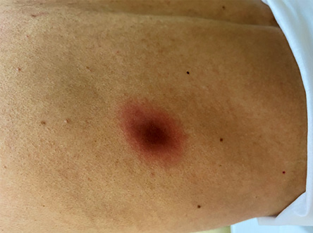 Figure 1: Solitary, edematous, oval erythematous plaque with a central duskier coloration on the skin of the left posterior chest.

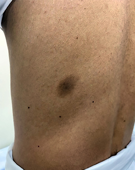 Figure 2: The site after clearance, showing post-inflammatory pigmentation.
Question
What is the most likely diagnosis?
a. Insect bite reaction.
b. Erythema multiforme.
c. Erythema migrans.
d. Fixed drug eruption.
Answer
d. Fixed drug eruption.
The diagnosis is fixed drug eruption (FDE) based on the history and typical skin lesion. Pseudoephedrine was suspected to be the culprit drug as previously when the patient took paracetamol alone no such skin lesions had occurred, while a rechallenge with pseudoephedrine hydrochloride alone resulted in a similar lesion on the same site. Naranjo adverse drug reaction probability scale was > 9; therefore, histopathology examination was not done. The patient was treated with topical steroids which resulted in clearance of the lesion leaving post-inflammatory pigmentation [Figure 2].
Discussion
FDE is an immunological cutaneous adverse drug reaction characterized by skin lesions that occur at the same location every time there is exposure to the causative substance.1 Many clinical variants have been described including pigmented, non-pigmented, generalized, linear, generalized bullous, urticarial, and oral.
FDE classically presents as a single, or a few, oval, sharply demarcated erythematous and edematous plaques with a dusky violaceous hue.2 There may be a central blister and associated pruritus or burning sensation. The most commonly affected sites are the trunk, limbs, lips, palms, soles, penis, and groin. The eruption appears one to two weeks after first exposure and within 24 hours of subsequent exposures.2
Many drugs can induce FDE. The most common drugs are paracetamol, tetracyclines, non-steroidal anti-inflammatory drugs, dapsone, quinine, aspirin, sulfonamides, and phenolphthalein.1–3 The ingestion of the causative agent may occur via any route, including oral, rectal, or intravenous. Pseudoephedrine hydrochloride is known to cause a non-pigmenting variant of FDE, however pigmenting variants caused by it have been reported as in our case.3
The diagnosis is based on its typical presentation and lesion morphology. Skin biopsy is usually not necessary unless the diagnosis is not clear. Systemic (oral) or topical (patch) provocation tests can be performed to identify the drug when the history is not clear or multiple drugs are suspected.4 Differential diagnoses include insect bite reaction, erythema multiforme, and erythema migrans. These diagnoses have no relation to drugs and lesions do not recur on the same site. The number of lesions, distribution, and morphology with target lesions help further differentiate erythema multiforme from FDE. Insect bite reaction is differentiated by history of insect bite and clinical appearance of a central punctum. Erythema migrans have a typical presentation of a bull’s eye-like lesion with slow expansion and associated history of a tick bite.
Avoidance of the offending drug is the most important step in the management of FDE. Topical steroids with or without oral antihistamines may be used to alleviate associated pruritus and erythema. Patients with generalized FDE can be treated with a short course of oral prednisolone 0.5–1 mg/kg per day for 3–5 days.5
Disclosure
The authors declared no conflicts of interest. Written consent was obtained from the patient.
Acknowledgments
We thank the patient for giving consent to publish the case.
references
- 1. Burgdorf WH, Plewig G, Wolff HH, Landthaler M, editor. 3rd ed. Berlin: Springer-Verlag; 2009. Braun Falco’s Dermatology. p. 460-462.
- 2. Bolognia JL, Jorizzo JL, Schaffer JV. Dermatology 3rd ed. Elsevier Health Sciences; 2012. p. 345.
- 3. Ozkaya E, Elinç-Aslan MS. Pseudoephedrine may cause “pigmenting” fixed drug eruption. Dermatitis 2011 May;22(3):E7-E9.
- 4. Shiohara T. Fixed drug eruption: pathogenesis and diagnostic tests. Curr Opin Allergy Clin Immunol 2009 Aug;9(4):316-321.
- 5. Patel S, John AM, Handler MZ, Schwartz RA. Fixed drug eruptions: an update, emphasizing the potentially lethal generalized bullous fixed drug eruption. Am J Clin Dermatol 2020 Jun;21(3):393-399.