An 85-year-old farmer presented with a painless, gradually progressing proliferative lesion on his left temporal scalp close to pinna. After ignoring it for 22 years, its unexpected rapid growth over the last month made him seek medical attention. It started as a small peanut size swelling that spontaneously discharged jelly-like whitish substance which encrusted over the years to give the present pedunculated appearance. There was no history of trauma, bleeding, or itching at the site. He had no addictions. His past and family histories were insignificant.
General physical and systemic examinations were within normal limits. Local examination revealed a 1 × 1 cm globular, non-tender, soft-tissue swelling on the left temporal scalp with two distinct horn-like structures bifurcating from it. It was free from the underlying structures [Figure 1]. There were no similar lesions elsewhere on the body. There was no associated significant peri-auricular or cervical lymphadenopathy, and the ipsilateral external ear examination did not reveal any abnormality.
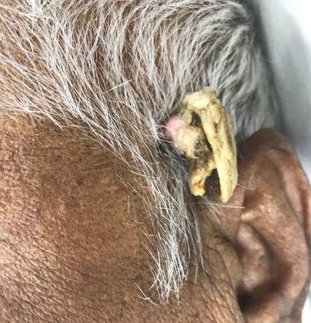
Figure 1: Keratotic dermal outgrowth. Note the nodular soft-tissue lesion with bifurcating pedunculated projections simulating a typical
animal horn.
The patient’s routine hemogram as well as x-rays of the skull and the chest were all normal. Mantoux test was negative. Thus, an excisional biopsy was performed under local anesthesia with a 1 cm margin all around and the wound was closed primarily. The microscopic evaluation revealed basal epidermal atypia and palisading of basal cells without any granulomas. Presence of solar elastosis suggested prolonged sun exposure. However, the squamous epithelial cells did not show pleomorphism or keratinization.
Question
What is the probable diagnosis?
a) Cutaneous tuberculosis.
b) Squamous cell carcinoma.
c) Cutaneous horn.
d) Giant viral wart.
Answer
c) Cutaneous horn.
The described clinico-pathological features are consistent with cutaneous horn with associated actinic keratosis. The patient recovered well postoperatively. He is under regular follow-up since last six months and has no recurrence.
Discussion
Cutaneous horns, also known as cornu cutaneum, are conical projections typically consisting of densely cohesive hyperkeratotic material with morphological resemblance to animal horns; however, without any bony core.1–4 Its earliest documentation dates to 1588 in an elderly Welsh woman.1–3,5 These lesions are frequently seen in sun-exposed areas like face, neck, hands, and forearm.3 Usually occurring after the fifth decade of life, they can vary in size, shape, and number.2 Cutaneous horns are rare among Asians and Africans, but common among Caucasians.5 One reason might be the protective nature of melanin pigment in the former. Though the exact reason remains elusive, chronic solar radiation exposure, immunodeficiency, and viral warts have been implicated in its pathogenesis.5
Over 60% of cutaneous horn lesions are benign.3 However, the possibility of an underlying malignancy should always be kept in mind while evaluating an elderly patient having a large wide-based cutaneous horn with tender, erythematous, ulcerated, or nodular base exhibiting fixity to the deeper structures, especially in presence of significant regional lymphadenopathy.1,2,5 Such cases mandate detailed evaluation using advanced imaging technologies.
Cutaneous horn has several close differential diagnoses; it needs a keen eye for their clinical distinctions, as follows: pilomatricoma usually presents in the first two decades of life as a subcutaneous lesion with a reddish blue hue on it; and pyogenic granuloma, a lobular capillary hemangioma, a rapidly growing benign lesion generally seen in young adults with history of local trauma, has a bright red appearance and bleeds easily on touch. Cutaneous tuberculosis has varied non-specific dermal presentations like papules, vesicles, or pustules with erythematous rash and matted lymphadenopathy. However, it should be underscored that the unique diagnostic feature of cornu cutaneum is its horn-like encrusted projections.
It is also important to know that a typically benign-looking corneum cutaneum may harbor a pre-malignant condition (commonest being actinic keratosis, as in this case) or even sub-clinical malignancy (commonest being squamous cell carcinoma).2,4,5 Though the diagnosis is primarily clinical, these issues potentiate its diagnostic enigma.3 Therefore, it is recommended that all cutaneous horns undergo full-thickness excision with adequate tumor-free margins of at least 1 cm, and subjected to dermato-pathological evaluation for better prognostication of the disease.1–5
references
- Singh P, Nathani D, Ranjan S, Issar R. A giant cutaneous horn projecting from verrucous carcinoma of buccal mucosa: a rare case report. J Clin Diagn Res 2017 Mar;11(3):ZD04-ZD05.
- 2. Soriano LF, Piansay-Soriano ME. A rapidly growing giant cutaneous horn on the chest. J Dermatol Case Rep 2015 Dec;9(4):113-115.
- 3. Copcu E, Sivrioglu N, Culhaci N. Cutaneous horns: are these lesions as innocent as they seem to be? World J Surg Oncol 2004 Jun;2(1):18.
- 4. Hermida Pérez JA, Bermejo Hernández A. Cuerno cutáneo, queratosis actínica y carcinoma espinocelular. A propósito de un caso clínico. Semergen 2013 Mar;39(2):113-116.
- 5. Gupta AK, Arora V, Gupta S, Bhagat T, Aggarwal A. Cutaneous horn — appearance may be deceptive. NJLM 2015 Apr;4(2):10-12.