Xanthogranulomatous cholecystitis (XGC) is rare benign inflammatory condition with a variable clinical presentation. Its etiology remains unknown, but its pathological features and variations are well known. However, reaching a preoperative diagnosis is extremely challenging due to its locally aggressive behavior. Additionally, tumor markers such as carbohydrate antigen 19-9 (CA19-9) might be elevated in this condition. Yet extremely high levels are usually attributed to biliopancreatic malignancy. However, there are three cases reported with extremely high preoperative CA19-9 levels that normalized after cholecystectomy.1 Moreover, there are reported cases of concomitant malignancy in patient with XGC.2 Hence, we present a puzzling clinical manifestation of XGC with extremely high CA19-9 levels without the presence of a malignancy component, contributing to the ongoing dilemma regarding the best management approach.
Case report
A 62-year-old man complained of episodes of colicky right upper quadrant pain with associated nausea, vomiting, and loss of appetite for the last two months. Recently, he developed a very painful episode with associated jaundice without itchiness. He was evaluated in the regional hospital and was found to have biochemical and radiological evidence of obstructive jaundice. He was referred to our institute for further work-up and management. On examination, his vital signs were normal. His abdominal examination revealed a fullness in the right upper quadrant with associated tenderness.
His laboratory investigation showed hemoglobin levels of 11.1 g/dL (NR: 11.5–15.5 g/dL), platelet count was 375 × 109/L (normal range (NR): 150–400 109/L), and the white cell count was 5.6 × 10⁹/L (NR: 4.5–11 × 109/L). His electrolytes and renal function were within normal limits. His liver enzymes were as follows: alanine aminotransferase 171 iU/L (NR: 7–55 iU/L), aspartate aminotransferase 131 iU/L (NR: 8–33 iU/L), and alkaline phosphatase 545 iU/L (NR: 44–147 iU/L). He had a normal albumin of 39 g/L (NR: 34–54 g/L) and a raised total bilirubin of 179 µmol/L (NR: 1.71–20.5 µmol/L). His coagulation was as follows: prothrombin time 12.7 s (NR: 10–13 s), activated partial thromboplastin time 39.7 s (NR: 30–40 s), and international normalized ratio of 1.16. His hepatitis screen was negative. His tumor markers profile was as follows: alpha fetoprotein 4.2 µg/L (NR: 0.5–15 µg/L), carcinoembryonic antigen 0 U/mL (NR: 0–2.5 U/mL), and CA19-9 2364 U/mL (NR: 0–37 U/mL).
Computed tomography (CT) scan demonstrated irregular diffuse mural thickening of the gallbladder wall, hypoattenuating mural nodules, multiple hyperdense calculi, and pericholecystic fluid collection, with poor fat planes to the adjacent liver parenchyma [Figure 1]. Abdominal magnetic resonance imaging (MRI) revealed a diffusely thickened gallbladder wall with iso- to slightly hyperintense signal, along with a few mural nodules within the thickened wall, indicating fat deposition of xanthogranuloma. Additionally, a focal area of early enhancement of adjacent liver parenchyma was observed [Figure 2].
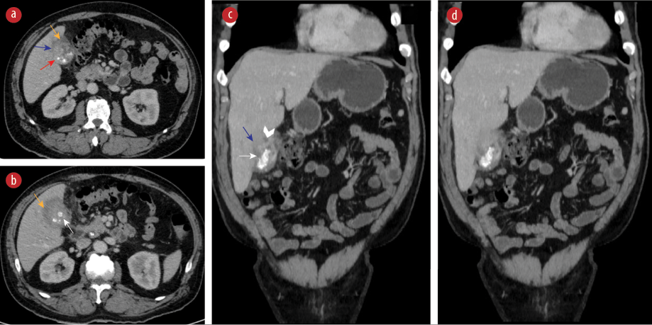 Figure 1: Contrast-enhanced CT scans of the abdomen in (a, b) coronal and (c, d) axial views demonstrate irregular diffuse mural thickening of the gallbladder wall (red arrow) along with few hypoattenuating mural nodules (white head arrows). Multiple hyperdense calculi (white arrows) and pericholecystic fluid collection (yellow arrows). Poor fat planes to the adjacent liver parenchyma are noted (blue arrows).
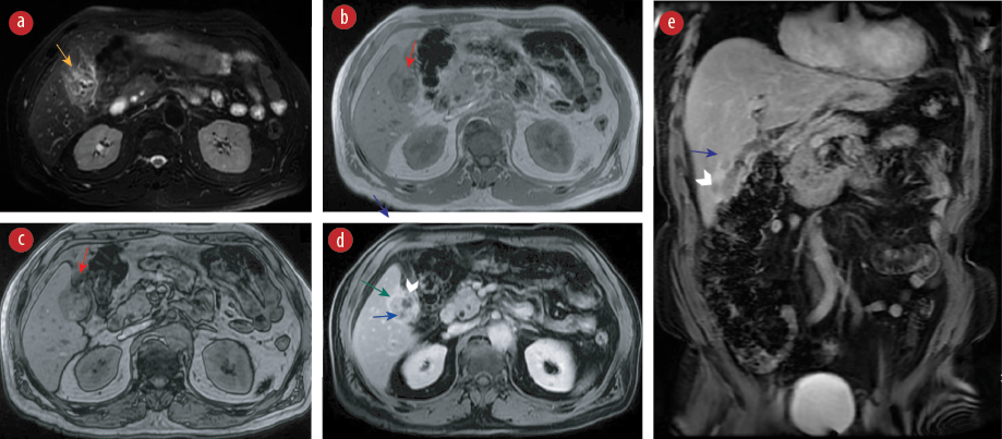 Figure 2: MRI of the abdomen including (a) axial fat-suppressed T2WI, (b) in-phase, (c) opposed-phase chemical shift images, (d) T1WI post contrast in axial and € coronal demonstrate a diffusely thickened gallbladder wall with iso- to slightly hyperintense signal in T2WI (yellow arrow) along with a few mural nodules within the thickened wall (white head arrows) some of which demonstrate signal drop out on opposed-phase images, indicating fat deposition of xanthogranuloma (red arrows). Minimal pericholecystic fluid is noted. Smooth luminal surface enhancement is seen on post-contrast images (blue arrows). A focal area of early enhancement of adjacent liver parenchyma is also seen (green arrow).
He was referred urgently to the gastroenterology for endoscopic retrograde cholangiopancreatography to decompresses his biliary system. Two weeks later, under conscious sedation, a successful endoscopic retrograde cholangiopancreatography was performed, including sphincterotomy, retrieval of common bile duct stone, and placement of a plastic stent. A week later, he was admitted for radical surgical resection. During admission, his total bilirubin level showed a significant reduction to 30 µmol/L. All his liver enzymes normalized, except for alkaline phosphatase, which significantly reduced to 184 iU/L. His coagulation parameters also normalized.
He underwent radical cholecystectomy with wedge resection of segment 4b and 5 along with porta hepatis lymph adenectomy via a right subcostal incision. During surgery, a segment of the greater omentum and the anterior wall of the hepatic flexure colon were resected as en bloc with the specimen. The cystic margin and frozen sections of the hepatic margins were negative for malignancy. He lost approximately 100cc of blood. His postoperative period was unremarkable, apart from a superficial wound infection.
Histopathology showed gallbladder wall exhibiting mucosal ulceration and dense infiltration by mixed inflammatory cell infiltrate, comprising mainly foamy histiocytes containing bile pigment admixed with multinucleated giant cells, lymphocytes, and plasma cells. Areas of marked fibrosis were seen with adherent liver tissues. There was no evidence of dysplasia or malignancy [Figure 3].
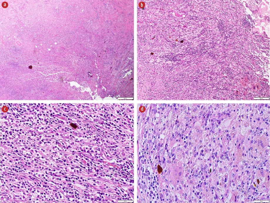 Figure 3: Histopathological slides of the gallbladder. (a) Histologic appearance of gallbladder wall showing mucosal ulceration and dense inflammation (magnification = 4 ×),(b) histologic appearance showing the dense inflammation with abundant histiocytes, multinucleated giant cells admixed with lymphoplasmacytic cell infiltrate (magnification = 10 ×), (c) showing abundant plasma cells (magnification = 40 ×), (d) demonstrating the details of the inflammatory cells (magnification = 40 ×).
On follow-up outpatient visit, he was doing well and asymptomatic. His CA19-9 level has normalized to 13 U/mL. He was discharged from the clinic with no further follow-up.
Discussion
XGC is a rare benign idiopathic gallbladder pathology that may simulate malignancy both radiologically and pathologically. It is characterized by marked increase in gallbladder wall thickness with lymphocytes and plasma cells in a focal or diffuse inflammatory process, which can extended into the adjacent structures such as the liver as in our case. The gross examination may reveal yellow masses within thickened gallbladder wall. Nevertheless, the definitive diagnosis can only be obtained by pathological examination.2
Xanthogranulomatous reaction secondary to inflammation is different from xanthomatous lesions cause by hypercholesterolemia. Cozzutto et al,3 in their review of the xanthogranulomatous process, postulated a hypothesis for the formation of foamy macrophages. They found that they are formed in medias where hemorrhage, suppuration, and necrosis occur profoundly and for long periods such as chronic or remittent severe inflammation. Basically, the macrophages lysosomes phagocytic activity cannot cope with the digestion of huge amount of destroyed blood cell by-products such as phospholipids.3
Although the imaging diagnosis of XGC is challenging, some characteristic imaging findings may suggest the diagnosis preoperatively.4 The main differential diagnoses of XGC are carcinoma of the gallbladder, actinomycosis, and adenomyomatosis of the gallbladder.5,6
On ultrasound, the main finding of XGC is marked mural thickening of the gallbladder with associated gallstones or sludge. Parra et al,5 noted that the wall thickening was hyperechoic compared with the liver parenchyma in all cases. Hypoechoic nodules or bands in the thickened wall can be seen in approximately 19% of XGC cases,5,7 and considered a characteristic finding. Ultrasound also can demonstrate associated complications such as perforation, abscess formation, and adjacent hepatic parenchymal infiltration.5,8
CT findings include diffuse or focal wall thickening, intramural hypodense nodules in thickened walls, luminal surface enhancement with continuous or focal disruption of mucosal line. Glabladder calculi and pericholecystic fluid collection are commonly associated with XGC.9 Infiltration into adjacent liver parenchyma or bowel can also be seen.9 CT may also demonstrate other complications such as perforation, abscess, or fistula formation.4,7
Ito et al,4 concluded that using CT scoring system, which assessed the presence of five components, including diffuse wall thickening, absence of polypoid lesions, intramural nodules or bands, pericholecystic infiltration, and pericholecystic abscess collection, likely increases the sensitivity and specificity of CT diagnosis of XGC. The presence of three or more findings had a sensitivity of 77% and specificity of 94% (with 95% CI). Zhao et al,9 studied the CT findings in 49 proven cases of XGC and found that diffuse wall thickening was seen in 87.8% and intramural hypodense nodules in the thickened wall were noted in 85.7% of cases. Luminal surface enhancement with continuous mucosal line was observed in 85.7% of cases. Gallbladder stones were seen in 69.4% of patients. These CT findings were observed in 40% of cases and 80% had four or more of these features. Coexistent malignancy of the gallbladder has been documented.6,10 Krishnani et al,10 found that 19.6% of XGC cases had associated carcinoma of the glabladder. In our patient, CT showed irregular diffuse glabladder mural thickening with a few hypoattenuating mural nodules, multiple hyperdense calculi, along with pericholecystic fluid collection and poor fat planes to the adjacent liver parenchyma.
MRI can add further characterization of XGC. In T2-weighted images, the thickened gallbladder wall with xanthogranuloma accumulation shows areas of iso- to slightly hyperintense signal, which show variable enhancement in post contrast study.11 Luminal surface enhancement of the glabladder wall denotes intact epithelial layer.7,9 Early enhancement of adjacent liver parenchyma indicates liver involvement. In-phase and opposed-phase chemical shift imaging is useful to detect the presence of fat within intramural nodules in XGC.11 This sign is likely highly specific and can lead to the diagnosis. Diffusion-weighted images helps to further differentiate between thickened glabladder wall in XGC and glabladder cancer. Kang et al,12 noticed that diffusion restriction was commonly observed in glabladder cancer (68%) compared with 7% of XGC cases. In our patient, MRI revealed a diffusely thickened glabladder wall, which shows iso- to slightly hyperintense signal on T2-wieghted images, along with few mural nodules within the thickened wall, some of which demonstrate signal dropout in opposed-phase images, denoting microscopic fat deposition of xanthogranuloma. Minimal pericholecystic fluid was seen. Smooth luminal surface enhancement is noted on post-contrast images. The presence of microscopic fat on MRI helped in reaching the diagnosis preoperatively.
CA19-9 is a cell surface glycoprotein complex produced by ductal cells from different gastrointestinal and non-gastrointestinal organs. Its expression and metabolism are affected by many factors, including enzyme deficiency, ethnicity, diet, and comorbidities. However, overexpression is known to occur more commonly with pancreaticobiliary pathology, specifically pancreaticobiliary malignancy. Carcinogenesis related epigenetic silencing factor is responsible for malignant overexpression of CA19-9, owing to considering it as a tumor marker. Nevertheless, the emerging terminology now is tumor-associated consequent to its significant association with vast benign pathology. Nonetheless, extremely high levels of CA19-9 are generally found in advance cases of pancreaticobiliary malignancy, although it is not exclusive.13,14
In begin biliary condition, acute cholangitis, choledocholithiasis, and cholecystitis are the most reported case associated with extremely high CA19-9 levels. This suggests that obstructive inflammation is probably the culprit for elevated CA19-9. Although the exact cause of extremely high CA19-9 levels in benign condition is unknown, there are few theories that have been postulated. The first is that the raised inflammatory cytokines lead to over expression of CA19-9. The second is that extensive inflammation leads to ductal obstruction and large amount of cellular destruction, leading to increase production and hematogenous leakage.14,15
In XGC, CA19-9 have played a significant role in assessment alongside clinical and radiological evaluation to distinguish between gallbladder adenocarcinoma and XGC. However, emerging evidence of extremely high CA19-9 levels seen in XGC, question its significance. Yu et al,16 have reported a raised CA19-9 in 45.95% of patients with XGC reaching a maximum 536.29 U/mL. On the other hand, Zhuang et al,17 studied the association of XGC to gallbladder carcinoma, and found that although CA19-9 is significantly higher in gallbladder carcinoma, it cannot be used to distinguish between them. The rise in CA19-9 levels in patients with XGC is yet not fully understood, as it may rise in the absence of jaundice. Furthermore, it is reported that the diseased gallbladder epithelium in XGC express CA19-9, signifying the rise is disease related.1
Conclusion
Although XGC is considered a benign disease, its complexity and variability require an aggressive approach in assessment. Likewise, the locally aggressive nature of the condition, as well as its association and similarity with gallbladder carcinoma, compels clinicians to take decisive measures in management to avoid suboptimal treatment.
Disclosure
The authors declare no conflicts of interest. Written consent was obtained from the patient.
references
- 1. Iida T, Adachi T, Nakagaki S, Yabana T, Goto A. A case of xanthogranulomatous cholecystitis with high CA19-9 levels that normalized post-cholecystectomy. J Clin Case Rep 2016;6(710):2.
- 2. Saritas AG, Gul MO, Teke Z, Ulku A, Rencuzogullari A, Aydin I, et al. Xanthogranulomatous cholecystitis: a rare gallbladder pathology from a single-center perspective. Ann Surg Treat Res 2020 Oct;99(4):230-237.
- 3. Cozzutto C, Carbone A. The xanthogranulomatous process. Xanthogranulomatous inflammation. Pathol Res Pract 1988 Aug;183(4):395-402.
- 4. Ito R, Kobayashi T, Ogasawara G, Kono Y, Mori K, Kawasaki S. A scoring system based on computed tomography for the correct diagnosis of xanthogranulomatous cholecystitis. Acta Radiol Open 2020;9(4):2058460120918237.
- 5. Parra JA, Acinas O, Bueno J, Güezmes A, Fernández MA, Fariñas MC. Xanthogranulomatous cholecystitis: clinical, sonographic, and CT findings in 26 patients. AJR Am J Roentgenol 2000 Apr;174(4):979-983.
- 6. Suzuki H, Wada S, Araki K, Kubo N, Watanabe A, Tsukagoshi M, et al. Xanthogranulomatous cholecystitis: difficulty in differentiating from gallbladder cancer. World J Gastroenterol 2015;21(35):10166-10173.
- 7. Casas D, Pérez-Andrés R, Jiménez JA, Mariscal A, Cuadras P, Salas M, et al. Xanthogranulomatous cholecystitis: a radiological study of 12 cases and a review of the literature. Abdom Imaging 1996;21(5):456-460.
- 8. Kim PN, Ha HK, Kim YH, Lee MG, Kim MH, Auh YH. US findings of xanthogranulomatous cholecystitis. Clin Radiol 1998 Apr;53(4):290-292.
- 9. Zhao F, Lu PX, Yan SX, Wang GF, Yuan J, Zhang SZ, et al. CT and MR features of xanthogranulomatous cholecystitis: an analysis of consecutive 49 cases. Eur J Radiol 2013 Sep;82(9):1391-1397.
- 10. Krishnani N, Shukla S, Jain M, Pandey R, Gupta RK. Fine needle aspiration cytology in xanthogranulomatous cholecystitis, gallbladder adenocarcinoma and coexistent lesions. Acta Cytol 2000;44(4):508-514.
- 11. Shuto R, Kiyosue H, Komatsu E, Matsumoto S, Kawano K, Kondo Y, et al. CT and MR imaging findings of xanthogranulomatous cholecystitis: correlation with pathologic findings. Eur Radiol 2004 Mar;14(3):440-446.
- 12. Kang TW, Kim SH, Park HJ, Lim S, Jang KM, Choi D, et al. Differentiating xanthogranulomatous cholecystitis from wall-thickening type of gallbladder cancer: added value of diffusion-weighted MRI. Clin Radiol 2013 Oct;68(10):992-1001.
- 13. Lee T, Teng TZ, Shelat VG. Carbohydrate antigen 19-9 - tumor marker: past, present, and future. World J Gastrointest Surg 2020 Dec 27;12(12):468-490.
- 14. Tsen A, Barbara M, Rosenkranz L. Dilemma of elevated CA 19-9 in biliary pathology. Pancreatology 2018 Dec;18(8):862-867.
- 15. Doğan ÜB, Gümürdülü Y, Gölge N, Kara B. Relationship of CA 19-9 with choledocholithiasis and cholangitis. Turk J Gastroenterol 2011;22(2):171-177.
- 16. Yu H, Yu TN, Cai XJ. Tumor biomarkers: help or mislead in the diagnosis of xanthogranulomatous cholecystitis?-analysis of serum CA 19-9, carcinoembryonic antigen, and CA 12-5. Chin Med J (Engl) 2013 Aug;126(16):3044-3047.
- 17. Zhuang PY, Zhu MJ, Wang JD, Zhou XP, Quan ZW, Shen J. Xanthogranulomatous cholecystitis: a clinicopathological study of its association with gallbladder carcinoma. J Dig Dis 2013 Jan;14(1):45-50.