A 10-year-old girl presented to the Emergency Department with severe epigastric pain, nausea, vomiting, and 15% weight loss. The vomiting was non-bloody, non-bilious, and associated with poor oral intake for two months. There were no associated loose stools, fever, or headache. Her medical history was significant for a vegan diet of two years; however, there was no history suggestive of body image issues.
Clinical examination revealed a thin girl with reduced muscle bulk. Her weight was 27 kg (5th centile), her height was 137 cm (25th centile), and her body mass index was 14 (5th centile). Her vital signs showed blood pressure of 104/75 mmHg, heart rate of 89 bpm, and a temperature of 36.7 oC. There was no orthostatic tachycardia or hypotension. Her abdominal examination revealed a scaphoid abdomen, which was soft with some epigastric tenderness. Her cardiopulmonary examinations were unremarkable.
Investigations showed hemoglobin of 10.4 g/dL (reference range: 11.5–15.5 g/dL) with mean corpuscular hemoglobin of 29 pg (24–33 pg), Mean corpuscular volume of 85.5 fL (73–95 fL), red cell distribution width of 14.5% (1.5–16.5%). White cell count and differential were normal. Inflammatory markers, liver enzymes, and renal function tests were also normal. Her thyroid function test, vitamin D, and lactate levels were also normal. Her bone profile showed alkaline phosphatase at 80 U/L (129–417 U/L), with normal albumin, calcium, and phosphate levels.
For evaluation of abdominal pain and vomiting, she underwent an upper gastrointestinal contrast study [Figure 1] and an abdominal computed tomography (CT) scan [Figure 2].
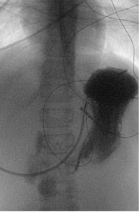 Figure 1: Upper gastrointestinal single-contrast study. Prolonged contrast hold-up was noted at the mid-duodenum. The stomach was partially decompressed by a nasogastric tube.
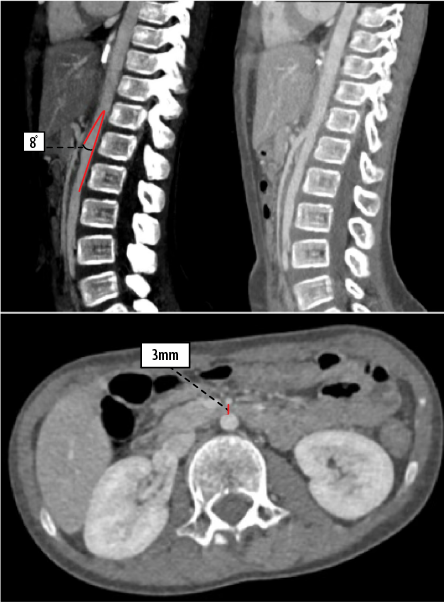 Figure 2: Contrast-enhanced CT of the abdomen and pelvis showing a reduced aortomesenteric angle measuring 8° and a reduced aortomesenteric distance of 3 mm. The third part of the duodenum is compressed. These measurements, along with fluoroscopic findings, are suggestive of superior mesenteric artery syndrome.
Questions
- What is the most likely diagnosis?
- What is the management of this condition?
Answers
- Superior mesenteric artery syndrome.
- Gastric decompression with a nasogastric tube, fluid and electrolyte correction, and nutritional rehabilitation, either enteral or parenteral, to promote weight gain.
Discussion
The upper gastrointestinal study demonstrated contrast hold-up at the level of the third part of the duodenum [Figure 1]. Abdominal CT showed a reduced aortomesenteric angle of 8 degrees and a reduced distance of 3 mm [Figure 2], consistent with a diagnosis of superior mesenteric artery syndrome (SMAS). SMAS, also known as Wilkie’s syndrome, is a rare condition that typically presents as mid- to upper gastrointestinal obstruction caused by an acquired vascular compression of the duodenum between the aorta and the SMA.1,2 Although it commonly affects young females in their twenties, it can occur at any age. Recent literature has also reported an increasing number of cases among elderly patients.3
SMAS in children is extremely rare, and its exact prevalence remains unknown due to the lack of pediatric-specific epidemiological studies. The prevalence is thought to be similar to that reported in the general population (0.01–0.3%).3 However, the incidence rises to 2–3% among children undergoing spinal surgery.4
SMAS is usually an acquired condition associated with significant weight loss or catabolic states, such as anorexia nervosa, severe burns, malabsorption syndromes, malignancy, and recent upper abdominal gastrointestinal surgeries. Mesenteric fat helps preserve this angle; however, with significant weight loss, the fat cushion diminishes, leading to a sharper angle and resulting in the SMA compressing the third part of the duodenum. This results in further weight loss, thereby perpetuating a vicious cycle.5
Additionally, patients undergoing scoliosis surgery may develop SMAS due to increased tension on the mesentery following spinal correction.3 However, congenitally short or hypertrophic ligament of Treitz, low origin of the SMA, an inherently narrow angle between the SMA and aorta, reduced aortomesenteric distance, and intestinal malrotation may also contribute to the development of SMAS in children.3
Patients with SMAS typically present with postprandial abdominal pain, persistent emesis, prolonged absence of stooling, and weight loss. They are often misdiagnosed with constipation. SMAS should be considered as a differential diagnosis, particularly in patients with acute weight loss.2,6 The diagnosis of SMAS requires a high index of clinical suspicion. In addition to the clinical picture, several imaging modalities can aid in diagnosis. Abdominal sonography may reveal a dilated duodenum with or without gastric dilatation. CT scan is considered the gold standard as it allows for measurement of the aortomesenteric angle and visualization of duodenal compression. The aortomesenteric angle is considered normal if it is between 28 and 65 degrees. The aortomesenteric distance is considered normal if it is between 10–34 mm.7 Upper gastrointestinal barium study may demonstrate contrast pooling in the stomach with delayed transit into the duodenum. Endoscopy can be used to detect gastric complications.3,8
Management of SMAS is typically conservative and focuses on bowel decompression, correction of the fluid and electrolyte imbalance, and nutritional support such as total parenteral nutrition. Once the patient is stabilized, small, frequent, high-calorie oral feeds are introduced to promote weight gain.1 Adjustment in feeding position, such as knees-to-chest or right lateral decubitus position, may also help alleviate the symptoms by facilitating duodenal passage of food.3 If symptoms persist, placement of a nasojejunal tube beyond the obstruction point may allow for continuous enteral feeding. When conservative management fails, particularly in the presence of profound proximal dilatation and stasis, surgical intervention becomes the second line. Surgical options include duodenojejunostomy, gastrojejunostomy, or resection of the ligament of Treitz.1
In this patient, following confirmation of the diagnosis, she was initially managed with a nasogastric tube for gastric decompression. Enteral nutrition was later initiated via a nasojejunal tube, resulting in a 7 kg weight gain during admission. She was transitioned to oral feeding and discharged home in a stable condition.
In summary, SMAS is a rare cause of duodenal obstruction in children. It is typically characterized by compression of the third part of the duodenum and a reduced aortomesenteric angle and distance. Clinicians should maintain a high index of suspicion for it, especially in the context of recent weight loss or surgery such as spinal correction. Management includes gastric decompression, fluid and electrolyte correction, and nutritional support. Early recognition and appropriate management are vital to improving outcomes and avoiding complications.
Disclosure
The authors declare no conflicts of interest.
references
- Biank V, Werlin S. Superior mesenteric artery syndrome in children: a 20-year experience. J Pediatr Gastroenterol Nutr 2006 May;42(5):522-525.
- 2. Waheed KB, Shah WJ, Jamal A, Mohammed HR, Altaf B, Amjad M, et al. Superior mesenteric artery syndrome: an often overlooked cause of abdominal pain! Saudi Med J 2021 Oct;42(10):1145-1148.
- 3. Oka A, Awoniyi M, Hasegawa N, Yoshida Y, Tobita H, Ishimura N, et al. Superior mesenteric artery syndrome: diagnosis and management. World J Clin Cases 2023 May;11(15):3369-3384.
- 4. Tsirikos AI, Jeans LA. Superior mesenteric artery syndrome in children and adolescents with spine deformities undergoing corrective surgery. J Spinal Disord Tech 2005 Jun;18(3):263-271.
- 5. Bozzola E, Irrera M, Cirillo F, Zanna V, Petrelli I, Diamanti A, et al. Superior mesenteric artery syndrome in anorexia nervosa: a case report and a systematic revision of the literature. Nutrients 2024 Feb;16(4):541.
- 6. England J, Li N. Superior mesenteric artery syndrome: a review of the literature. J Am Coll Emerg Physicians Open 2021 Jun;2(3):e12454.
- 7. Lamba R, Tanner DT, Sekhon S, McGahan JP, Corwin MT, Lall CG. Multidetector CT of vascular compression syndromes in the abdomen and pelvis. Radiographics 2014;34(1):93-115.
- 8. Kim JY, Shin MS, Lee S. Endoscopic features for early decision to evaluate superior mesenteric artery syndrome in children. BMC Pediatr 2021 Sep;21(1):392.