Osteoid osteoma ranks as the third most prevalent benign bone tumor, accounting for 10–14% of all benign bone tumors and 2–3% of primary bone tumors. It occurs predominantly in the long bones of the appendicular skeleton and specifically in the lower extremities.1 It is usually seen in school-aged children and adolescents.2 It is uncommon in children under five, constituting only 3% of cases.3
It usually presents with localized nocturnal pain that is alleviated by nonsteroidal anti-inflammatory drugs.3 Other symptoms are variable and depend on the location of the osteoid osteoma. However, in younger age groups, presentations can vary, potentially delaying diagnosis.4 The symptoms can include pain and limping when the lesion affects the lower extremities or can mimic osteomyelitis.4,5
Neurological manifestations can be the presenting symptoms in patients affected by this benign tumor.6 In this report, we describe a patient with right humerus osteoid osteoma who presented with right arm weakness. This weakness was associated with localized muscle wasting and depressed deep tendon reflexes on the affected side.
Case Report
A four-year-old male, with an uneventful perinatal history and appropriate development, presented with right-sided weakness persisting for almost three months. The parents first noticed that the child did not move his right arm when running. This progressed to noticeable floppiness. The patient also started complaining of pain in his arm that was partially relieved by massaging the proximal arm. Furthermore, the inability to move the right arm progressed, and the patient could not raise his right arm above 90 degrees above his shoulder. Moreover, he started waking up from sleep screaming from pain. There was no involvement of the lower extremities, ocular, bulbar, or facial muscles. Moreover, no further trauma and/or infectious symptoms were reported.
Clinical examination revealed a hypotonic right upper arm with a decrease in muscle bulk that involved the entire arm, predominantly the proximal arm muscles. Muscle strength examination showed 4/5 strength in shoulder abduction, elbow extension, elbow flexion, wrist extension, finger extension, and extensor indicis. In contrast, full power was obtained in the flexor digitorum profundus and the flexor digitorum superficialis. At that point, the working diagnosis was a lower motor neuron lesion, and from a localization perspective, the prospective brachial plexus was the first consideration.
Nerve conduction studies (NCS) and electromyography (EMG) were performed. The NCS showed low compound muscle action potentials involving ulnar, radial, musculocutaneous, and axillary nerves with normal distal latencies and conduction velocities. Sensory nerves were normal with no evidence of involvement. A needle exam performed on the right deltoid showed no spontaneous activity and normal motor units. This picture did not fit with the brachial plexus pattern. A possible plexopathy was the working diagnosis. Magnetic resonance imaging (MRI) of the brachial plexus and cervical spine was performed and showed no compressive lesions or enhancements in the cervical spine or brachial plexus. However, a focal area of signal abnormality was incidentally seen in the diaphysis of the right humerus with adjacent edema in the muscle as it was imaged partially and could not be further characterized on that MRI.
An X-ray of the right humerus showed a well-defined centric, slightly expansile, mixed density lesion in the diaphysis of the midshaft of the humerus with evidence of mild cortical resorption with no periosteal reaction, no soft tissue component, and no cortical disruption [Figure 1]. The patient was referred to a pediatric orthopedic surgeon for evaluation of the right humerus lesion identified on the X-ray. MRI and computed tomography findings were suggestive of a benign lesion of chondroid origin [Figure 2]. Therefore, a computed tomography-guided biopsy was taken, and the result confirmed the diagnosis of osteoid osteoma. After six months of treatment with radiofrequency ablation and physiotherapy, the child regained full right arm strength (5/5), and no muscle wasting was noted. Written consent was obtained from the patient’s father.
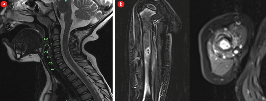 Figure 1: (a) MRI of the cervical spine, including brachial plexus, was normal, and (b) the humerus showing an enhancing lesion at the midshaft.
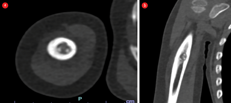 Figure 2: Axial and coronal CT sections showing a lesion at the mid-diaphysis of the humerus with thickened periosteum.
Discussion
Our patient presented with right arm weakness associated with generalized muscle atrophy. These symptoms were subsequently followed by the classic pain that is usually associated with osteoid osteoma. It is not uncommon for patients diagnosed with osteoid osteoma to present with neurological symptoms.3 Indeed, neurological symptoms can be the first manifestation of osteoid osteoma and can manifest prior to pain, which is considered a cardinal feature of this benign bone tumor.3 Keirs et al,6 reported seven pediatric patients with a confirmed histopathological diagnosis of osteoid osteoma involving the femur or tibia. All patients reported having localized muscle atrophy and/or depressed deep tendon reflexes. Moreover, another seven patients from the same cohort presented initially with neurological symptoms of abnormal gait and muscle wasting prior to their diagnosis. They were all evaluated by pediatric neurologists prior to their diagnosis.6 In addition, one patient from the cohort with osteoid osteoma involving the proximal humerus presented with palpable swelling leading to compression of the brachial plexus on the same side.6 Our patient had no brachial plexus compression, as confirmed by brachial plexus MRI. The osteoid osteoma was not in close proximity to the brachial plexus to suggest a possible explanation for the compressive lesion causing right arm weakness; however, that was the first working diagnosis for this patient. The NCS and EMG finging did not fit the brachial plexus injury pattern, and this was further supported by the normal brachial plexus MRI, which showed no evidence of any compressive lesions. Moreover, around 30% of patients with osteoid osteoma in the cervical spine can present with neurological deficits.3 However, the osteoid osteoma in the case of this patient was not at the cervical spine. The symptoms of wasting and inability to move the right arm were likely related to the osteoid osteoma lesion itself and not to a compressive lesion on nerves or plexus. Pain probably played a role in the avoidance of movement on the affected arm, leading to the finding of muscle atrophy due to deconditioning and the impression of arm weakness. The underlying mechanism of depressed deep tendon reflexes and hypotonia remains unclear, possibly related to deconditioning or a focal myopathic process.
Additionally, there was no delay in the osteoid osteoma diagnosis of this patient; however, what was challenging was the right-arm weakness associated with his clinical presentation. In this report, muscle weakness was the initial and main complaint reported by the parents, followed by nocturnal pain at the time of presentation. There was significant muscle atrophy in the muscles of the right arm. The first report of muscle atrophy in osteoid osteoma was in 1979 in a four-and-a-half-year-old girl with left tibial osteoid osteoma.7 She was found to have atrophy of 2 cm of the left thigh and calf muscles with diminished left limb power.7 In 1993, Kaweblum et al,8 reported that 50% of their patient cohort with lower limb osteoid osteoma had muscular atrophy, and one in five patients had a leg length discrepancy. A retrospective review (from 1985 and 2000) reported that of 48 patients with upper extremity osteoid osteoma, 46 reported persistent pain, and the other two only presented with swelling.9 Muscle weakness with atrophy has not been reported in this cohort.10 NCS and EMG in our patient showed low compound muscle action potentials in all motor nerves tested in the right arm, with sparing of the sensory nerves. This was attributed to muscle atrophy. Needle examination did not support a neurogenic pattern. In addition, MRI of the cervical spine and brachial plexus showed no evidence of injury or any enhancement indicating inflammation. Therefore, brachial plexus involvement was excluded. Brachial plexus involvement can either be due to an acquired inflammatory process or a hereditary neuropathic process. Furthermore, follow-up a year after radiofrequency ablation for osteoid osteoma showed a normal exam with complete resolution of muscle atrophy and full strength.
Conclusion
This case underscores the importance of considering osteoid osteoma in pediatric patients presenting with atypical neurological symptoms. The rarity of muscle weakness and atrophy associated with this benign tumor highlights the need for a comprehensive evaluation, incorporating non-neurological factors such as pain, in the diagnostic approach. Understanding the diverse presentations of osteoid osteoma is crucial for accurate and timely diagnosis, particularly in challenging age groups.
Disclosure
The authors declare no conflicts of interest.
references
- 1. Zhang Y, Rosenberg AE. Bone-forming tumors. Surg Pathol Clin 2017 Sep;10(3):513-535.
- 2. Kitsoulis P, Mantellos G, Vlychou M. Osteoid osteoma. Acta Orthop Belg 2006 Apr;72(2):119-125.
- 3. Tepelenis K, Skandalakis GP, Papathanakos G, Kefala MA, Kitsouli A, Barbouti A, et al. Osteoid osteoma: an updated review of epidemiology, pathogenesis, clinical presentation, radiological features, and treatment option. In Vivo 2021;35(4):1929-1938.
- 4. Laliotis N, Chrysanthou C, Konstantinidis P, Papadopoulou L. Osteoid osteoma in children younger than 3 years of age. Case Rep Orthop 2019 Sep;2019:8201639.
- 5. Bhat I, Zerin JM, Bloom DA, Mooney JF III. Unusual presentation of osteoid osteoma mimicking osteomyelitis in a 27-month-old infant. Pediatr Radiol 2003 Jun;33(6):425-428.
- 6. Kiers L, Shield LK, Cole WG. Neurological manifestations of osteoid osteoma. Arch Dis Child 1990 Aug;65(8):851-855.
- 7. Black JA, Levick RK, Sharrard WJ. Osteoid osteoma and benign osteoblastoma in childhood. Arch Dis Child 1979 Jun;54(6):459-464.
- 8. Kaweblum M, Lehman WB, Bash J, Strongwater A, Grant AD. Osteoid osteoma under the age of five years. The difficulty of diagnosis. Clin Orthop Relat Res 1993 Nov:(296):218-224.
- 9. Themistocleous GS, Chloros GD, Benetos IS, Efstathopoulos DG, Gerostathopoulos NE, Soucacos PN. Osteoid osteoma of the upper extremity. A diagnostic challenge. Chir Main 2006 Jun;25(2):69-76.
- 10. Kaweblum M, Lehman WB, Bash J, Grant AD, Strongwater A. Diagnosis of osteoid osteoma in the child. Orthop Rev 1993 Dec;22(12):1305-1313.