A six-year-old boy with congenital achalasia cardia, who had undergone Heller myotomy and fundoplication 18 months earlier, presented with a two-week history of recurrent vomiting after food and fluid intake. The episodes progressively increased in frequency and severity. Initially, intolerance was limited to solid foods, but it later extended to all foods and liquids. To evaluate the cause of his dysphagia, the patient underwent esophagogastroduodenoscopy, which revealed inflamed esophageal mucosa with ulceration and whitish and yellowish plaque-like lesions in the lower third of the esophagus [Figure 1]. Written consent was obtained from the patient’s parents.
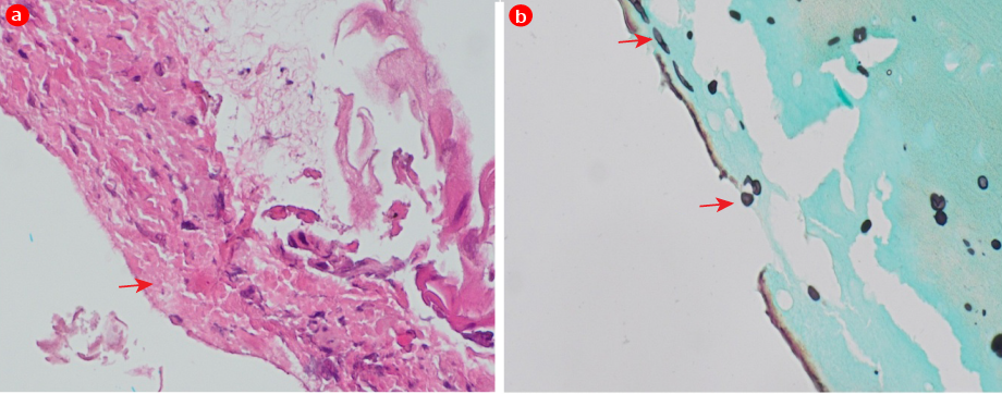 Figure 1: Endoscopy showing inflamed esophageal mucosa of the lower third of esophagus with whitish and yellowish plaque-like lesions, typical of Candida esophagitis.
Questions
- What is the diagnosis?
- What are the possible causes of vomiting after Hellar myotomy?
- How would you confirm the diagnosis?
- How would you manage this condition?
Answers
- The diagnosis is Candida esophagitis. The endoscopic image reveals inflamed esophageal mucosa with whitish and yellowish plaque-like lesions, typical of Candida esophagitis.
- Possible differential diagnosis: (a) Incomplete myotomy – obstruction may persist because the myotomy did not fully divide the lower esophageal sphincter. (b) Tight fundoplication – too tight a wrap may obstruct the esophagogastric junction. (c) Recurrent or persistent achalasia – due to incomplete relief of the primary motility disorder. (d) Gastroesophageal reflux disease – may occur after myotomy due to loss of lower esophageal competence, leading to esophagitis and secondary dysphagia.
- The diagnosis can be confirmed by biopsy and/or brush cytology of the esophageal mucosa, which usually shows yeasts and pseudohyphae invading the mucosal cells. Fungal culture of the specimen may also identify Candida.
- The condition is managed with systemic antifungal therapy, typically with fluconazole.
Discussion
Histological examination of esophageal mucosa showed inflammation with focal ulceration, Candida spores, and pseudohyphae [Figure 2]. The child was treated with oral fluconazole for two weeks and the family subsequently reported improvement in symptoms. He currently experiences occasional vomiting with solid foods, which represents his baseline due to the underlying motility disorder.
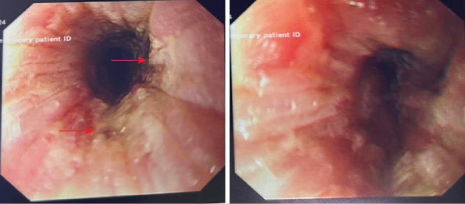 Figure 2: (a) Hematoxylin and eosin-stained section and (b) grocott methenamine silver-stained section, both at × 40 magnification, showing fungal pseudohyphae in the esophageal biopsy.
Esophageal achalasia (EA) is a rare esophageal motor disorder affecting all age groups, caused by an abnormal absence of ganglion cells in the Auerbachian muscle layer in the lower esophagus. EA rarely occurs in children below five years of age. Symptoms occur because of high resting tension and impaired relaxation of the lower esophageal sphincter.1
The main objective in the management of EA is to reduce the pressure of the lower esophageal sphincter to facilitate normal peristalsis. Heller myotomy with a partial anti-reflux procedure is considered the gold standard due to its high effectiveness and safety. More recently, peroral endoscopic myotomy has been introduced as a minimally invasive technique.2
Infectious esophagitis occurs predominantly in immunocompromised patients.3 Young children are less commonly affected.3 The principal pathogens are Candida species, herpes simplex virus, and cytomegalovirus. Candida esophagitis is typically associated with impaired immunity due to malignancy or immunosuppressive therapy.3 In a recent Romanian study of 520 children undergoing upper gastrointestinal endoscopy for symptoms such as dysphagia, heartburn, and appetite loss, post-procedure infectious esophagitis was identified only in 11 (2.1%) children, including five (1.0%) cases of Candida esophagitis. Among the infectious esophagitis, Candida was the most common etiology (5; 45.5%), followed by cytomegalovirus (4; 36.4%) and herpes simplex virus (2; 18.2%).3 The study identified immunodeficiency (81.8%) and prolonged broad-spectrum antibiotic therapy as the principal risk factors for infectious esophagitis.3
The diagnostic criteria for esophageal candidiasis include its characteristic endoscopic appearance with white or slightly yellowish plaque-like lesions on the esophageal mucosa, together with microscopic evidence of pseudohyphae in endoscopic mucosal brushings or invasive candidiasis on biopsy.4
Although Candida esophagitis is uncommon in patients with achalasia, literature reviews and isolated case reports have suggested an association.5 Esophageal stasis due to impaired peristalsis may lead to retention of food and secretions, mucosal maceration, and altered pH, thereby predisposing to secondary opportunistic infections, with Candida being the most frequently encountered pathogen.5 Evidence linking achalasia to Candida esophagitis is derived primarily from adult-focused studies. A large retrospective cohort of 234 patients with achalasia (median age = 45 years) reported a 12% prevalence of esophageal Candida infection, markedly higher than the 0.3–5.2% estimated in the general population; however, the study did not provide age-stratified data to clarify pediatric involvement.5 Although infectious esophagitis commonly affects immunocompromised patients, esophageal stasis and poor clearance due to motility disorders such as achalasia predisposes even immunocompetent individuals to Candida infection, which is considered as the most common infectious cause of dysmotility-associated esophagitis.
Esophageal candidiasis generally responds to systemic antifungal therapy, with oral fluconazole as the preferred first-line agent. Intravenous fluconazole is used when oral dosing is not feasible. Alternatives include itraconazole and voriconazole, while amphotericin B and posaconazole are reserved for refractory or severe disease. Management follows standard pediatric infectious esophagitis approaches; no achalasia-specific pediatric guidelines are available.4 Although adult studies suggest an association with increased esophageal cancer risk, pediatric outcomes are unknown.5 Infectious esophagitis may cause notable morbidity, especially in immunocompromised children, but most cases improve with timely antifungal therapy.3
Conclusion
Although Candida esophagitis is uncommon in children, it should be considered in pediatric patients with esophageal dysmotility including achalasia and post-Heller status who present with recurrent vomiting or dysphagia. Esophageal stasis and impaired clearance of food can predispose even immunocompetent children to secondary Candida infection.
Disclosure
The authors declare no conflicts of interest.
references
- 1. Marlais M, Fishman JR, Fell JM, Haddad MJ, Rawat DJ. UK incidence of achalasia: an 11-year national epidemiological study. Arch Dis Child 2011 Feb;96(2):192-194.
- 2. Jarzębicka D, Czubkowski P, Sieczkowska-Gołub J, Kierkuś J, Kowalski A, Stefanowicz M, et al. Achalasia in children—clinical presentation, diagnosis, long-term treatment outcomes, and quality of life. J Clin Med 2021 Aug;10(17):3917.
- 3. Bordea MA, Pîrvan A, Gheban D, Silaghi C, Lupan I, Samașca G, et al. Infectious esophagitis in Romanian children: From etiology and risk factors to clinical characteristics and endoscopic features. J Clin Med 2020 Mar;9(4):939.
- 4. Mohamed AA, Lu X-L, Mounmin FA. Diagnosis and Treatment of Esophageal Candidiasis: Current Updates. Can J Gastroenterol Hepatol 2019 Oct;2019:3585136.
- 5. Guo X, Lam SY, Janmaat VT, de Jonge PJ, Hansen BE, Leeuwenburgh I, et al. Esophageal Candida Infection and Esophageal Cancer Risk in Patients With Achalasia. JAMA Netw Open 2025 Jan;8(1):e2454685.