Infantile hemangiomas (IHs), occurring in 4–12% of infants, represent the most frequent benign tumors in this age group.1,2 PHACES syndrome (posterior fossa malformations, hemangioma, arterial anomalies, coarctation of the aorta/cardiac defects, eye abnormalities, and sternal malformations), classified as a neuro-cutaneous syndrome, was initially identified by Frieden and colleagues in 1996.3 This syndrome is observed in 2% of patients with segmental hemangiomas on the cervicofacial distribution, particularly in the frontotemporal or maxillary/mandibular regions. This condition is associated with various anomalies, including posterior fossa abnormalities, facial hemangiomas, arterial cerebrovascular anomalies, cardiac anomalies, coarctation, ocular anomalies, and sternal defects.4,5 The arteriopathy accompanying PHACES syndrome poses a significant concern in the treatment of patients with high-risk vascular anomalies who may require propranolol therapy. This arteriopathy carries the potential to initiate ischemic strokes, making it a primary focus in patient management. It is essential to exercise caution when considering propranolol therapy for individuals with high-risk vascular anomalies associated with PHACES syndrome, as there is a possibility that the use of propranolol could increase the risk of stroke in such cases. We describe the clinical and radiological characteristics of three patients with PHACES syndrome who exhibited cerebrovascular arteriopathy. Our focus is on the treatment challenges and clinical outcomes. The clinico-radiological findings of the three cases are summarized in Table 1.
Table 1: Summary of the clinico-radiological findings of the three cases.
|
Age, weeks
|
3
|
5
|
2
|
|
Gender
|
Male
|
Male
|
Female
|
|
Location of hemangioma
|
Head and neck
|
Face
|
Face
|
|
Extra-cutaneous hemangioma
|
Nil
|
posterior paraspinal muscles from C1-C3
|
Intestinal
intracranial
|
|
Eye
|
Normal
|
Normal
|
Optic disc anomaly
|
|
Cardiac
|
Normal
|
Normal
|
Normal
|
|
Arteriopathy
|
Absence of the right internal carotid artery
|
Left internal carotid artery has a smaller caliber compared to the right side
|
Hypoplastic right internal carotid artery
|
Written consent for publication was obtained from all patients' parents.
Case Report
Case one
A three-week-old late preterm neonate presented with a segmental infantile hemangioma that affected the right neck, face, and scalp. The extent of the hemangioma raised suspicion of PHACES syndrome [Figure 1]. A magnetic resonance angiogram (MRA) was performed using a time-of-flight technique and revealed a congenital absence of the right internal carotid artery (ICA) in both cervical and intracranial segments, without any significant intracranial abnormalities [Figure 1]. The patient underwent ophthalmology and cardiology evaluations, which showed no abnormalities. A magnetic resonance imaging (MRI) perfusion study was attempted but was unsuccessful due to technical limitations. The multidisciplinary team decided to monitor the patient since he was asymptomatic and had a high stroke risk. During follow-up, the patient's hemangioma showed minimal growth with no complications. As a prophylactic measure, aspirin was initiated to prevent stroke.
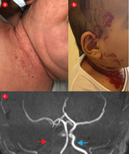 Figure 1: Segmental infantile hemangioma involving the right side of the (a) neck, (b) right face, and right scalp, which progressed over time as shown in the pictures. (c) Time-of-flight MRA of the intracranial vessels shows absent of the right intracranial internal carotid artery (red arrow), including its cervical segments (not shown here), compared with the normal left internal carotid artery (blue arrow).
Case two
An 11-week-old male infant born at term presented with progressive right facial swelling at the age of five weeks. The main presenting symptom was noisy breathing, without any stridor or shortness of breath. Clinical examination revealed a well-circumscribed, soft, 5 cm right cheek swelling with telangiectatic changes [Figure 2]. Ultrasound imaging revealed a hypervascular heterogeneously hypoechoic lesion over the right cheek consistent with a high-flow soft tissue hemangioma. Given the segmental appearance of the hemangioma, PHACES syndrome was suspected. MRI of the head and neck showed a right cheek mass and multiple other lobular lesions in the posterior paraspinal muscles from C1-C3, all of which had signal characteristics and an enhancement pattern consistent with hemangiomas [Figure 2]. No evidence of extension into the spinal canal was noted. MRA revealed that the left internal carotid artery had a smaller caliber compared to the right side, with focal mild to moderate stenosis of the left internal carotid artery just distal to the carotid bifurcation. The vertebral arteries had a mildly tortuous course. Cardiac and ophthalmology assessments were normal. The decision to initiate propranolol was debatable due to the high risk of stroke associated with intracranial arterial anomalies, leading to a multidisciplinary discussion that ultimately preferred to proceed with sclerotherapy as the best option for this infant. Several challenges delayed the intervention, given the patient's age and weight, including the unavailability of a small catheter size. Therefore, the team decided to start low-dose propranolol (1 mg/kg/day) while awaiting sclerotherapy. The infant showed a good response and improvement in symptoms while on low-dose propranolol, which was eventually tapered and discontinued after the right cheek lesion had regressed during follow-up at 12 months of age.
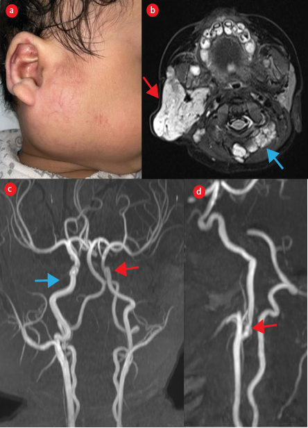 Figure 2: (a) Right cheek swelling with some telangiectatic changes over it. (b) Axial T2-weighted images with fat saturation shows hemangiomas in the right parotid space (red arrow) and posterior paraspinal muscles (blue arrow). (c) Maximum intensity projection of time-of-flight MRA shows a smaller caliber of the left internal carotid artery (red arrow) compared with the right internal carotid artery (blue arrow). (d) Focal stenosis of the proximal left internal carotid artery just distal to the carotid bifurcation (arrow).
Case three
A three-month-old female neonate born at full-term presented with segmental hemangioma, which first appeared as a telangiectatic patch on the right periorbital area and forehead at two weeks of age. The lesion progressed to redness and swelling over time [Figure 3]. A thorough evaluation for PHACES syndrome revealed hypoplasia of the right internal carotid artery via MRA [Figure 3], an optic disc anomaly, and normal cardiac assessment. During the same hospitalization, she developed intussusception and required laparotomy after unsuccessful pneumatic reduction. The intussusception was attributed to an ileum hemangioma, which was excised. Given the presence of hemangiomas in three critical sites (i.e., orbital, intracranial, and intestinal hemangioma), a low dose of oral propranolol at 1.5 mg/kg/day was initiated. The treatment was effective, allowing for the gradual tapering and eventual cessation of propranolol by 24 months of age.
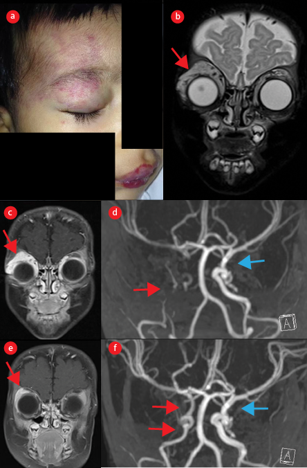 Figure 3: (a) Telangiectatic patch over the right periorbital, forehead, and lips, which then progressed to redness and swelling. (b) Coronal T2-weighted image with fat suppression and (c) post-contrast T1- weighted image, at the age of three months, show the right orbital hemangioma (arrows). (d) Time-of-flight MRA of the proximal intracranial arteries shows absence of the right internal carotid artery (red arrow) compared to the normal left internal carotid artery (blue arrow). (e) At the age of six months, after treatment with propranolol, the right orbital hemangioma shows an interval reduction in size (arrow), with (f) marked improvement in recanalization of the right internal carotid artery (red arrows), although it remains smaller in caliber compared to the left internal carotid artery (blue arrow).
Discussion
PHACES syndrome is a rare disorder of unknown etiology and pathogenesis. The extracutaneous manifestations of this syndrome are well-documented and include cerebrovascular anomalies, which are associated with potential comorbidities such as seizures and ischemic strokes.4,6 The documented arteriopathy predominantly affects the medium and large-caliber cerebral vessels, giving rise to stenosis, occlusion, agenesis, hypoplasia, or anomalous origin/course of the main cerebral arteries, saccular aneurysms, and arterial dysplasia.7,8 Furthermore, intracranial vasculopathy such as Moyamoya syndrome may cause arterial ischemic stroke.5,9
Patients with PHACES syndrome who present with significant narrowing (> 25%) or occlusion, aplasia or hypoplasia of main cerebral vessels, tandem or multiple arterial stenoses that diminish cerebral perfusion, and/or imaging findings suggesting chronic or silent brain ischemia are considered to be at high risk for arterial ischemic stroke.10,11 In this context, initiating of treatment for segmental hemangioma, which often occurs within the first few months of life, is crucial given its aggressive proliferation, and propranolol treatment is considered the gold standard therapy for visual impairment or airway obstruction.10,11
However, the presence of vasculopathy in children affected by PHACES syndrome raises concerns about the potential increased risk of stroke associated with beta-blocker use, especially in those with major or multiple arterial anomalies.12 While arterial anomalies are commonly observed in PHACES syndrome cases, there is limited information on the clinical outcomes of these changes after propranolol use. Two patients have reported stroke while receiving propranolol. The hypotension-induced reduction in blood flow in the stenotic, occluded, hypoplastic, or absent artery is the proposed mechanism by which propranolol may trigger ischemic stroke.12
Several reported cases of PHACES syndrome associated with strokes, with the average age of stroke occurrence being 13.6 months, and the most common presenting symptoms being seizure and hemiparesis, emphasize the challenges of starting oral propranolol in patients with arterial anomalies and the need for careful consideration of the potential risk of stroke.5
Our three cases highlight the multifaceted nature of propranolol therapy decisions in PHACES syndrome, emphasizing the importance of meticulous case evaluation and a multidisciplinary approach to treatment decision-making. In case one, the decision not to administer propranolol and instead opt for clinical observation was based on the assessment that the patient had a notable stroke risk, while the hemangioma did not induce functional abnormalities. For cases two and three, the high indication for propranolol therapy was driven by the potential for functional abnormalities arising from the hemangioma's location, coupled with a lower vascular stroke risk compared to case 1.
In summary, propranolol remains a key therapeutic option for managing segmental hemangioma in PHACES syndrome, but its use must be approached with caution, especially in patients with significant arterial anomalies. The potential risk of stroke underscores the importance of thorough evaluation and a multidisciplinary approach to treatment decision-making.
Conclusion
This case series involving three PHACES syndrome patients brings to light the challenging task of deciding on propranolol administration in those with cerebrovascular anomalies due to the increased susceptibility to stroke. It emphasizes the paramount importance of interdisciplinary discussions. Regrettably, the utility of brain perfusion study to assess cerebral blood flow before propranolol initiation as a potential predictor of stroke risk remains unclear. Additional research is imperative to determine the role of perfusion studies in the algorithm for propranolol administration in
these patients.
Disclosure
The authors declared no conflicts of interest.
References
- 1. Tekes A, Koshy J, Kalayci TO, Puttgen K, Cohen B, Redett R, et al. S.E. Mitchell vascular anomalies flow chart (SEMVAFC): a visual pathway combining clinical and imaging findings for classification of soft-tissue vascular anomalies. Clin Radiol 2014 May;69(5):443-457.
- 2. Hoornweg MJ, Smeulders MJ, Ubbink DT, van der Horst CM. The prevalence and risk factors of infantile haemangiomas: a case-control study in the Dutch population. Paediatr Perinat Epidemiol 2012 Mar;26(2):156-162.
- 3. Frieden IJ, Reese V, Cohen D. PHACE syndrome. The association of posterior fossa brain malformations, hemangiomas, arterial anomalies, coarctation of the aorta and cardiac defects, and eye abnormalities. Arch Dermatol 1996 Mar;132(3):307-311.
- 4. Metry DW, Haggstrom AN, Drolet BA, Baselga E, Chamlin S, Garzon M, et al. A prospective study of PHACE syndrome in infantile hemangiomas: demographic features, clinical findings, and complications. Am J Med Genet A 2006 May;140(9):975-986.
- 5. Metry D, Heyer G, Hess C, Garzon M, Haggstrom A, Frommelt P, et al; PHACE Syndrome Research Conference. Consensus statement on diagnostic criteria for PHACE syndrome. Pediatrics 2009 Nov;124(5):1447-1456.
- 6. Metry DW, Dowd CF, Barkovich AJ, Frieden IJ. The many faces of PHACE syndrome. J Pediatr 2001 Jul;139(1):117-123.
- 7. Heyer GL, Dowling MM, Licht DJ, Tay SK, Morel K, Garzon MC, et al. The cerebral vasculopathy of PHACES syndrome. Stroke 2008 Feb;39(2):308-316.
- 8. Heyer GL, Millar WS, Ghatan S, Garzon MC. The neurologic aspects of PHACE: case report and review of the literature. Pediatr Neurol 2006 Dec;35(6):419-424.
- 9. Garzon MC, Epstein LG, Heyer GL, Frommelt PC, Orbach DB, Baylis AL, et al. PHACE syndrome: consensus-derived diagnosis and care recommendations. J Pediatr 2016 Nov;178:24-33.e2.
- 10. Leboulanger N, Cox A, Garabedian EN, Denoyelle F. Infantile haemangioma and β-blockers in otolaryngology. Eur Ann Otorhinolaryngol Head Neck Dis 2011 Nov;128(5):236-240.
- 11. Siegel DH, Tefft KA, Kelly T, Johnson C, Metry D, Burrows P, et al. Stroke in children with posterior fossa brain malformations, hemangiomas, arterial anomalies, coarctation of the aorta and cardiac defects, and eye abnormalities (PHACE) syndrome: a systematic review of the literature. Stroke 2012 Jun;43(6):1672-1674.
- 12. Drolet BA, Frommelt PC, Chamlin SL, Haggstrom A, Bauman NM, Chiu YE, et al. Initiation and use of propranolol for infantile hemangioma: report of a consensus conference. Pediatrics 2013 Jan;131(1):128-140.