An eleven-month-old boy presented to the ophthalmology clinic with a history of strabismus and severe visual impairment of the right eye. There was no history of seizures, vomiting, loss of consciousness, or neurological deficits. He was developmentally appropriate for his age. Family history was noncontributory, with no history of neurofibromatosis type 1 or similar conditions. On examination, there were no dysmorphic features or skin changes. His neurological examination
was unremarkable.
Ophthalmic examination revealed a large angle esotropia in the right eye. The patient did not fixate with this eye. Anterior segment examination showed a central, dot-like opacity on the posterior surface of the lens. Dilated fundus examination revealed a large dysplastic optic nerve with peripapillary pigmentation and limited retinal elevation. A radial distribution of vessels and a tuft of glial tissue on the optic disc were noted [Figure 1]. A stalk of glial tissue extended from the central part of the optic disc to the posterior surface of the lens, identified as Cloquet’s canal, a remnant of fetal vasculature.
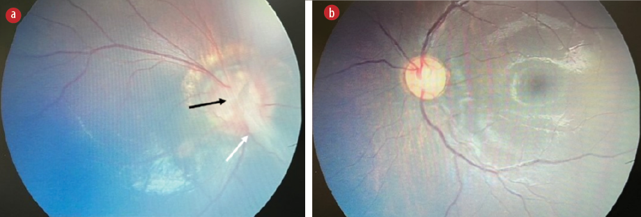 Figure 1: Color fundus photographs of the (a) right eye showing a large dysplastic optic nerve with peripapillary pigmentary disturbance. The retinal vessels are radially distributed on the optic disc. Note the tuft of glial tissue on the optic disc (black arrow) and a fibroglial stalk (white arrow) extending anteriorly from the center of the optic disc. This is a remnant of the Cloquet’s canal. The macula is hypoplastic. (b) The fundus photograph of the left eye shows a normal optic disc, macula, vessels, and retinal background.
Examination of the left eye revealed a persistent pupillary membrane in the anterior segment. Posterior segment examination was unremarkable. Magnetic resonance imaging (MRI) of the brain and orbit revealed a funnel-shaped excavation of the right optic disc associated with an elevated adjacent retinal surface and discontinuity of the uveoscleral surface. A thin, faint band of tissue was seen extending from the posterior surface of the lens to the optic nerve head along the Cloquet's canal, consistent with the fundoscopic findings. There was an absence of fluid signal intensity within the right optic nerve sheath around the right optic nerve [Figure 2]. Focal thickening and faint T2 signal intensity of the ipsilateral optic chiasm were noted without abnormal enhancement [Figure 3]. There was no evidence of other midline abnormalies. Magnetic resonance angiography was unremarkable and there was no evidence of Moyamoya phenomenon.
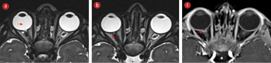 Figure 2: (a) Axial T2-sampling perfection with application optimized contrasts using different flip angle evolutions (SPACE) image shows funnel-shaped excavation of the right optic disc associated with elevated adjacent retinal surface and discontinuity of the uveoscleral surface (white arrow). A thin and faint band of tissue extending from the posterior surface of the lens to the head of the optic nerve along the Cloquet's canal (red arrow). (b) Axial T2-SPACE image demonstrates effacement of the fluid signal in the right optic nerve sheath around the right optic nerve (arrow). (c) Axial contrast-enhanced T1-weighted image with fat saturation shows marginal enhancement of the distal right optic nerve (arrow).
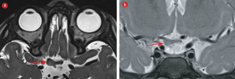 Figure 3: (a) Axial T2-sampling perfection with application optimized contrasts using different flip angle evolutions (SPACE) image and (b) coronal T2-weighted image with fat saturation show focal thickening and faint T2 hyperintensity of the ipsilateral optic chiasm (arrows).
Informed consent was obtained from the
patient’s parent.
Question
What is the diagnosis?
a. Coloboma.
b. Peripapillary staphyloma.
c. Morning glory disc anomaly.
d. Optic disc pit.
Answer
c. Morning glory disc anomaly.
Discussion
Morning glory disc anomaly (MGDA) is a rare congenital anomaly of the optic disc with a typical fundoscopic and radiological appearance.1,2 It manifests as a funnel-shaped excavation of the optic disc with chorioretinal pigmentary changes and radially oriented vessels resembling a morning glory flower.3 Patients may initially present with reduced visual acuity, leukocoria, and strabismus.3,4 Other ocular abnormalities that are described with MGDA include hypertelorism, persistent fetal vasculature, retinal detachment, congenital cataract, and drusen.3,5
Visual pathway abnormality is an important association of MGDA and includes ipsilateral thickening and contralateral hypoplasia of the optic chiasm secondary to axonal degeneration.4–7
Although previous literature on MGDA has labeled the observed abnormal thickening of the optic pathway as optic pathway glioma, recent studies considered this finding a developmental malformation associated with MGDA and should not be confused with optic pathway glioma.4,8,9 For instance, in a large retrospective study that included 32 patients with MGDA, 21 had mild segmental thickening of the ipsilateral optic nerve.8 Similarly, Nguyen et al,4 reported nine confirmed cases of MGDA. In eight of the cases, the thickness was variable and irregular along the optic pathway, including thick, thin, and normal segments. Serial MRI scans redemonstrated optic nerve abnormalities with no evidence of progression or evolution.
It is worth mentioning that in our patient, the thickened ipsilateral optic chiasm also demonstrated mild T2 hyperintensity, which is not a consistent feature in previously reported cases. In contrast, Firouzabadi et al,8 who studied a large cohort of patients with MGDA and optic pathway thickening, reported normal T2 signal intensity of the optic nerves and optic chiasm in all subjects.8 Therefore, a follow-up MRI has been planned for our patient.
MGDA is commonly associated with other facial and midline abnormalities, necessitating further brain and orbital MRI.1,2 Examples of midline abnormalities include cleft lip and palate, basal encephalocele, and corpus callosum agenesis.1,3 Other reported associated midline anomalies include persistent craniopharyngeal canal, pituitary gland and infundibular deformity, and tubular or nodular nasopharyngeal lesions.8 Intracranial vascular abnormalities, such as segmental aplasia or hypoplasia of the circle of Willis vessels and Moyamoya phenomenon, have also been described with MGDA.1,3,10
Although the majority of MGDA cases are unilateral, cases with bilateral MGDA have been reported in the literature.8,9 In addition, despite most cases of MGDA being sporadic, there are a few cases that were found to be familial and associated with other genetic mutations or other syndromes like PHACE syndrome, which is characterized by combinations of congenital anomalies, including posterior fossa malformations, hemangiomas, arterial anomalies, coarctation of the aorta and cardiac anomalies, and eye abnormalities.1
The cardinal MRI features of MGDA are funnel-shaped morphology of the posterior optic disc with elevation of the retinal surface, defect and discontinuity of the uveoscleral coat, and abnormal tissue associated with the ipsilateral distal intra-orbital segment of the optic nerve.2,4 These findings differentiate MGDA from other differential diagnoses and ocular anomalies such as coloboma and peripapillary staphyloma.2 Coloboma is a term used to describe any focal discontinuity in the structure of the globe.2,3 MGDA was described in some literature as a focal subtype of coloboma of the optic nerve head.2 Staphyloma occurs due to choroidal thinning and stretching without a defect.2 MGDA is also differentiated from coloboma and staphyloma by the presence of an abnormal central glial tuft, pigmentary changes, and radially oriented vessels, which are best seen during fundoscopic examination.1,3 Optimization of visual acuity with refractive correction and occlusion therapy of the fellow eye is crucial to prevent amblyopia.3
MGDA is a rare disc anomaly that has a typical fundoscopic and radiological appearance. Orbital and brain MRI is recommended in such cases as a screening tool to look for other midline anomalies and vascular abnormalities.
Disclosure
The authors declare no conflicts of interest.
references
- 1. Harasymowycz P, Chevrette L, Décarie JC, Hanna N, Aroichane M, Jacob JL, et al. Morning glory syndrome: clinical, computerized tomographic, and ultrasonographic findings. J Pediatr Ophthalmol Strabismus 2005;42(5):290-295.
- 2. Ellika S, Robson CD, Heidary G, Paldino MJ. Morning glory disc anomaly: characteristic MR imaging findings. AJNR Am J Neuroradiol 2013 Oct;34(10):2010-2014.
- 3. Lee BJ, Traboulsi EI. Update on the morning glory disc anomaly. Ophthalmic Genet 2008 Jun;29(2):47-52.
- 4. Nguyen DT, Boddaert N, Bremond-Gignac D, Robert MP. Optic nerve abnormalities in morning glory disc anomaly: an MRI study. J Neuroophthalmol 2022 Jun;42(2):199-202.
- 5. Kinori M, Smiley NP, Zeid JL. Morning glory disc anomaly and ipsilateral sporadic optic pathway glioma. Am J Ophthalmol Case Rep 2018;10:16-17.
- 6. Doneda C, Pinelli L, Scaramuzzi M, Galli J, Fazzi E, Parazzini C, et al. Morning glory disc anomaly associated with ipsilateral optic nerve and chiasm thickening: three cases and review of the literature. Neuropediatrics 2017 Dec;48(6):463-466.
- 7. Bandopadhayay P, Dagi L, Robison N, Goumnerova L, Ullrich NJ. Morning glory disc anomaly in association with ipsilateral optic nerve glioma. Arch Ophthalmol 2012 Aug;130(8):1082-1083.
- 8. Firouzabadi FD, Soldatelli MD, Rameh V, Heidary G, Vargas SO, Gonzalez E, et al. Morning glory disc anomaly: expanding the MR phenotype. AJNR Am J Neuroradiol 2024 Aug;45(8):1070-1075.
- 9. Ní Leidhin C, Erickson JP, Bynevelt M, Lam G, Lock JH, Wang G, et al. (What’s the story) morning glory? MRI findings in morning glory disc anomaly. Neuroradiology 2024 Jul;66(7):1225-1233.
- 10. Lenhart PD, Lambert SR, Newman NJ, Biousse V, Atkinson DS Jr, Traboulsi EI, et al. Intracranial vascular anomalies in patients with morning glory disk anomaly. Am J Ophthalmol 2006 Oct;142(4):644-650.