|
Introduction
A 50-year-old male presented with recurrent effusion around the left knee joint. There was no history of trauma to the knee or recent episode of fever. On clinical examination, suprapatellar fullness with decreased range of movement was noted. However, there was no tenderness or redness of the overlying skin. There were no complaints in other joints of the body or a similar family history. He was advised X-ray of the knee joint (anteroposterior and lateral view) which showed advanced degenerative changes in the form of reduced tibiofemoral joint space, osteophyte formation with subchondral sclerosis, and cystic changes. A large suprapatellar fullness was also seen (Fig. 1). He was advised magnetic resonance imaging (MRI) to look for any synovial or intraarticular pathology (Figs. 2, 3 and 4).
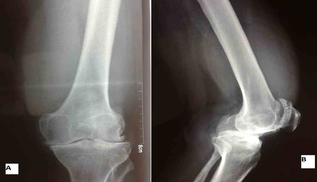
Figure 1: X-ray Left knee joint shows advanced degenerative changes with increased suprapatellar soft tissue density.

Figure 2: Axial and Sagittal T2 weighted MRI (A,B) shows suprapatellar effusion with frond-like synovial masses. Coronal T1 weighted MRI (C) shows hyperintense masses suggesting fatty origin.
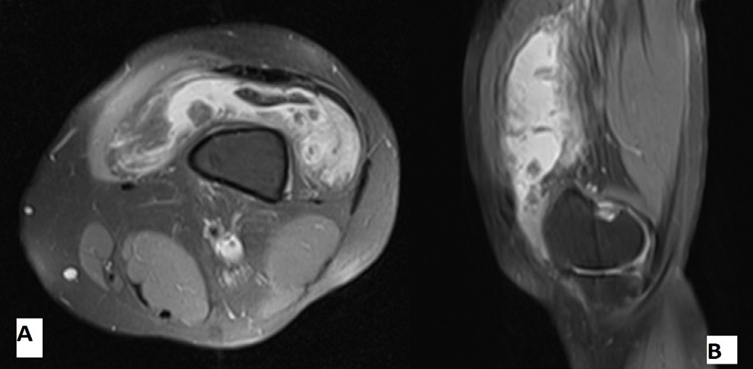
Figure 3: Fat suppressed Axial and Sagittal MRI confirms the fatty nature of synovial masses as they get suppressed while effusion does not.
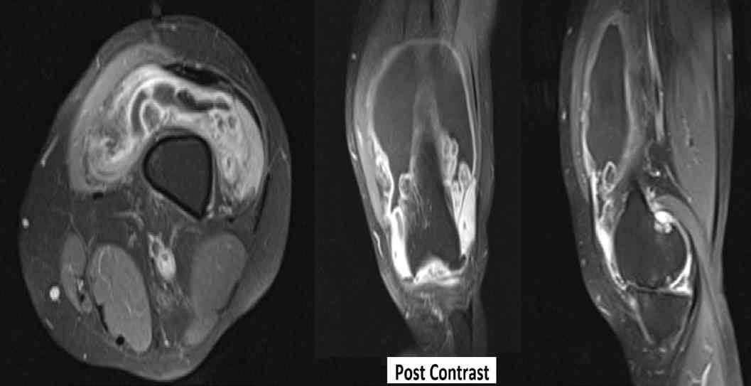
Figure 4: Gadolinium enhanced Axial, Coronal and Sagittal T1 weighted fat-suppressed MRI shows no enhancement of the fatty synovial mass but enhancement of the effusion.
Questions
1. What are the MRI findings and the role of MRI in this case?
2. What are the possible differentials in this case?
3. What is the line of management?
To access the full article, please complete the quiz online
|