|
Introduction
A 42-year-old male nonsmoker presented with recurrent hemoptysis for 12 years requiring two hospitalizations and 11 units of blood transfusion, with occasional low-grade intermittent fever, and cough with scanty expectoration. Examination of lower respiratory system revealed biphasic and coarse crepitations over the left infrascapular area.
Investigations revealed the following: Hb=75 gm/L; platelet count=250 × 109/L; prothrombin time=16.2 sec; INR=1.26; blood biochemistry was normal; sputum AFB was negative on 3 occasions. On USG, the whole abdomen was normal. A chest X-ray (Fig. 1) was also done. Subsequently, contrast-enhanced CT scans of thorax were done (Figs. 2 and 3). Finally, aortography was done. (Fig. 4)
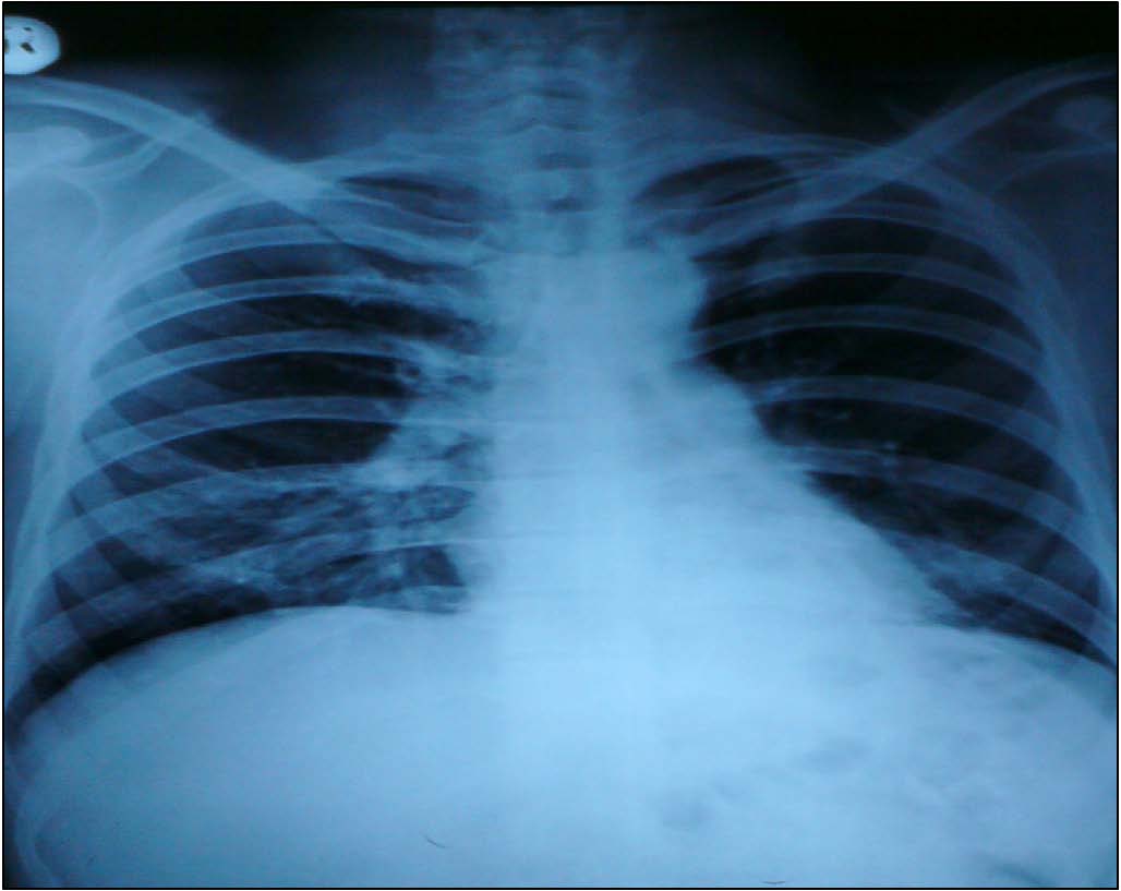
Figure 1: Chest X-ray was Normal.
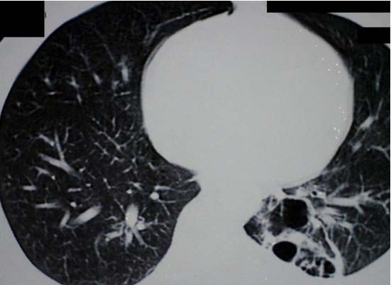
Figure 2: CECT Thorax (lung window) - Segmental collapse consolidation with cavity formation, breakdown, and associated bronchiectasis in posterior segment of left lower lobe.
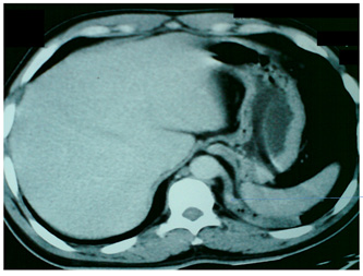
Figure 3: CECT Thorax (mediastinal window) - Lesion has separate blood supply of systemic circulation from descending thoracic aorta (arrow).
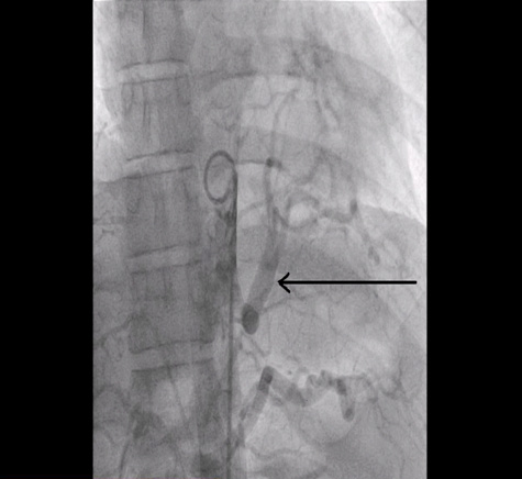
Figure 4: Aortography of Left lower lobe receiving a separate blood supply from the descending thoracic aorta (arrow).
Question
What is the diagnosis?
|