|
Introduction
A 69-year-old man visited our clinic with complaints of dyspnea. He had previously been diagnosed with dilated cardiomyopathy and prescribed a diuretic, angiotensin converting enzyme inhibitor, amiodarone (200 mg/day) and a ß-blocker for two years. A chest X-ray revealed pleural effusion and blood chemistry data revealed mildly elevated transaminases (AST 75 IU/L, ALT 95 IU/L). Our diagnosis was heart failure, and treatment was started with diuretics. However, even after amelioration of the heart failure, transaminase levels continued to be high at approximately 80 IU/L. Therefore, abdominal ultrasonography and computed tomography (CT) were performed to identify the cause, (Figs. 1, 2). The patient's laboratory data for iron, viral markers, and autoimmune markers were normal.
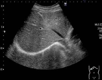
Figure 1. Ultrasonography.
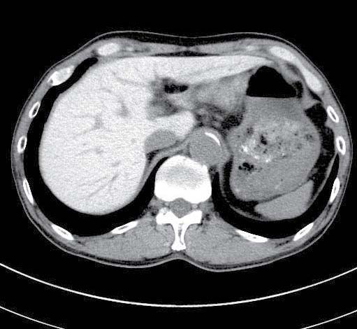
Figure 2. Computed Tomography.
Questions
- What are the abdominal ultrasonography and CT scan findings?
- What is your diagnosis?
- What are your treatment options?
Answers
1. Abdominal ultrasonography findings are normal but CT scan shows marked hyperdensity in the liver.
2. Liver dysfunction due to accumulation of amiodarone which contains iodine.
3. If possible, reduce the dose of amiodarone.
Discussion
Drug-induced liver dysfunction is very common. Therefore, when liver dysfunction is mild, we tend to neglect it. Recently however, amiodarone-induced liver dysfunction has been reported to be very dangerous. Indeed, some cases like this one have been reported and warnings have been given that follow-up only is far from adequate because there is some possibility of progression to cirrhosis.1,2 We have often recognized mild dysfunction as in this case in the past. However, abdominal ultrasonography alone was performed and findings were normal, such that we decided to continue with just follow-up. This is extremely risky. Now, it is believed that cases such as this one are not rare and that CT must be performed to actively prevent progression to cirrhosis. In fact, amiodarone dose reduction resulted in gradual improvement of transaminases in this case.
Acknowledgements
The authors reported no conflict of interest and no funding was received for this work.
References
1. Stravitz RT, Sanyal AJ. Drug-induced steatohepatitis. Clin Liver Dis 2003 May;7(2):435-451.
2. Grieco A, Forgione A, Miele L, Vero V, Greco AV, Gasbarrini A, et al. Fatty liver and drugs. Eur Rev Med Pharmacol Sci 2005 Sep-Oct;9(5):261-263.
|