|
Abstract
Ectrodactyly, also known as Split-Hand/Split-Foot Malformation (SHFM) is a rare genetic condition characterized by defects of the central elements of the autopod. It has a prevalence of 1:10,000-1:90,000 worldwide. The X-linked and autosomal dominant types have been described. It can occur as an isolated malformation or in combination with other anomalies, such as tibial aplasia, craniofacial defects, and genitourinary abnormalities. Ectrodactyly-ectodermal dysplasia-clefting syndrome (EEC) is an example of ectrodactyly syndrome accompanied by multiple organ defects. Ectrodactyly has been reported in Africa, especially in several families in remote areas of central Africa but there has not been any published work on ectrodactyly in Nigeria. A baby was born in Ilorin, North Central Zone of Nigeria, with an uneventful prenatal and delivery history but was noticed to have malformation of the two hands and the two lower limbs at birth which are replica of the father’s malformation. We present this case to highlight familial ectrodactyly in Nigeria and prepare us to improve upon simple prenatal diagnosis and management of the challenges associated with patients with congenital malformation in Nigeria and other developing countries.
Keywords: Familial; Ectrodactyly; Congenital.
Introduction
Ectrodactyly is a congenital limb malformation characterized by a deep median cleft of the hand and/or foot.1,2 The word ectrodactyly was coined from two Greek words - ektroma (abortion), and daktylos (finger).2 It is a rare congenital anomaly in Nigeria though the true prevalence is unknown. A case seen in our institution (University of Ilorin teaching Hospital, Ilorin, Kwara state, Nigeria) necessitated a literature review and a report.
Ectrodactyly was first documented in 1770 among a tribe of some Guiana in India.3 Other names for ectrodactyly include split-hand/foot malformation (SHFM), crab-claw deformity - first used by VonWalter in 1829, and lobster claw - first used by Cruveilhier in 1842.1 Some authors use ectrodactyly to denote any absence deformity of distal limbs and reserve SHFM for the typical malformation, while other use ectrodactyly synonymously with SHFM.4 The basic embryologic abnormality in ectrodactyly is failure to maintain a normal functioning apical ectodermal ridge, leading to failure of differentiation of the autopod (hand or foot).4 In ectrodactyly, the hand and/or foot appear split into two halves with failure of development of the phalanx, metacarpal and or metatarsal bones of one or more fingers and or toes. The spectrum of expression can be in form of just cutaneous syndactyly of digits to absence of the entire autopod,2,4,5 and several instances of non-penetrance have been documented.4
Ectrodactyly occurs in two forms, either as an isolated disorder or as a component of a syndrome. Any of these two forms can be sporadic or familial, sporadic cases being more common.4 The most frequent pattern of inheritance is autosomal dominant with reduced penetrance. Autosomal recessive6 and X-linked forms of transmission,7 are less frequent, while other cases of ectrodactyly are caused by chromosomal deletion and duplication.4 Common anomalies associated with ectrodactyly include tibial aplasia, craniofacial defects, and genitourinary abnormalities. Ectrodactyly-ectodermal dysplasia-clefting syndrome (EEC) is the prototypical example of ectrodactyly syndrome accompanied by multiple organ defects. It is defined by a triad of ectrodactyly, ectodermal dysplasia and cleft lip and palate.1,2 This is a rare genetic disorder with an incidence of 1:90,000.1,8 Other syndromes associated with ectrodactyly include Carpenter’s syndrome, DeLange syndrome, Goltz syndrome, and Miller syndrome.1
Ectrodactyly is said to be relatively common in few central African communities with possible common progenitor.9 These are namely; two groups from eastern Botswana and south-western Zimbabwe belonging to the Talaunda tribe and have a common ancestral origin. Another family are members of the Wadoma tribe of north-eastern Zimbabwe (described as the two-toed ostrich-foot people).9,10 Recent studies have found three families of affected individuals among these tribes.1 In Nigeria, this is the first case to be reported to the best of our knowledge.
Case Report
A 2 hour old term male neonate presented with a history of malformation of limbs noticed at birth. The mother had antenatal care at a basic health (referral) centre, where she booked at a gestational age of 12 weeks. No maternal illness in pregnancy and no fever or skin rash were noted. She had only routine drugs without a history of ingestion of any other medication or herbal concoction. No maternal smoking or alcohol ingestion was reported. Labour lasted four hours and delivery was a spontaneous vaginal delivery at the referral centre. The baby cried immediately after birth and passed meconium shortly after birth, and also while being attended to. This was the mother’s fourth child, but the second surviving child. The first child was an 8 year old boy who was well, the second and third children were also males but died at 18 and 14 months respectively from febrile illnesses. No history of similar malformations in any sibling. However, the father had similar isolated malformation in the limbs. Hence, there was no history of exposure to radiation in both parents. The father is a 36 year old driver, while mother is a 25 year old hairdresser, both with secondary school education. There was no history of consanguinity or any other congenital malformation. Examination findings revealed an otherwise active neonate with malformation of the upper and lower limbs. (Figs. 1-3)
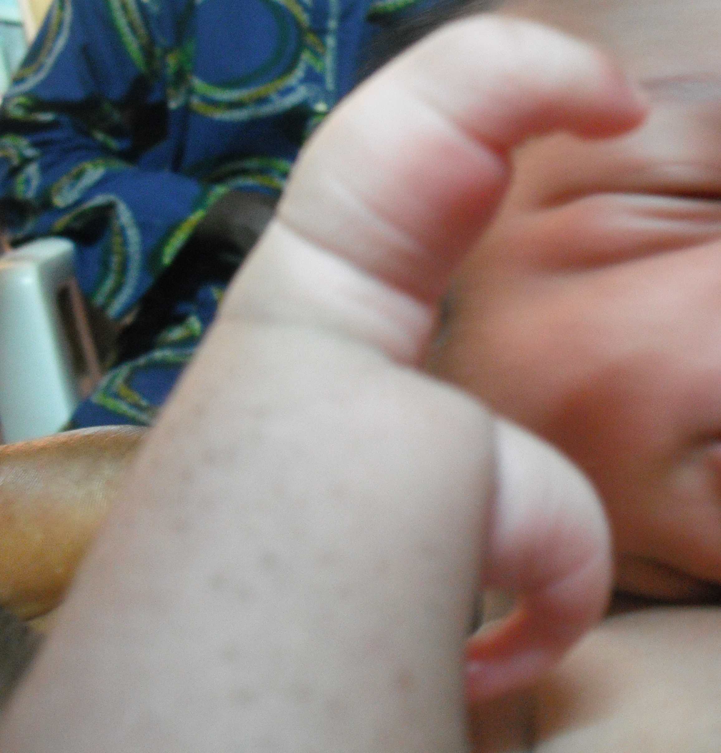
Figure 1: Baby’s Right Hand.
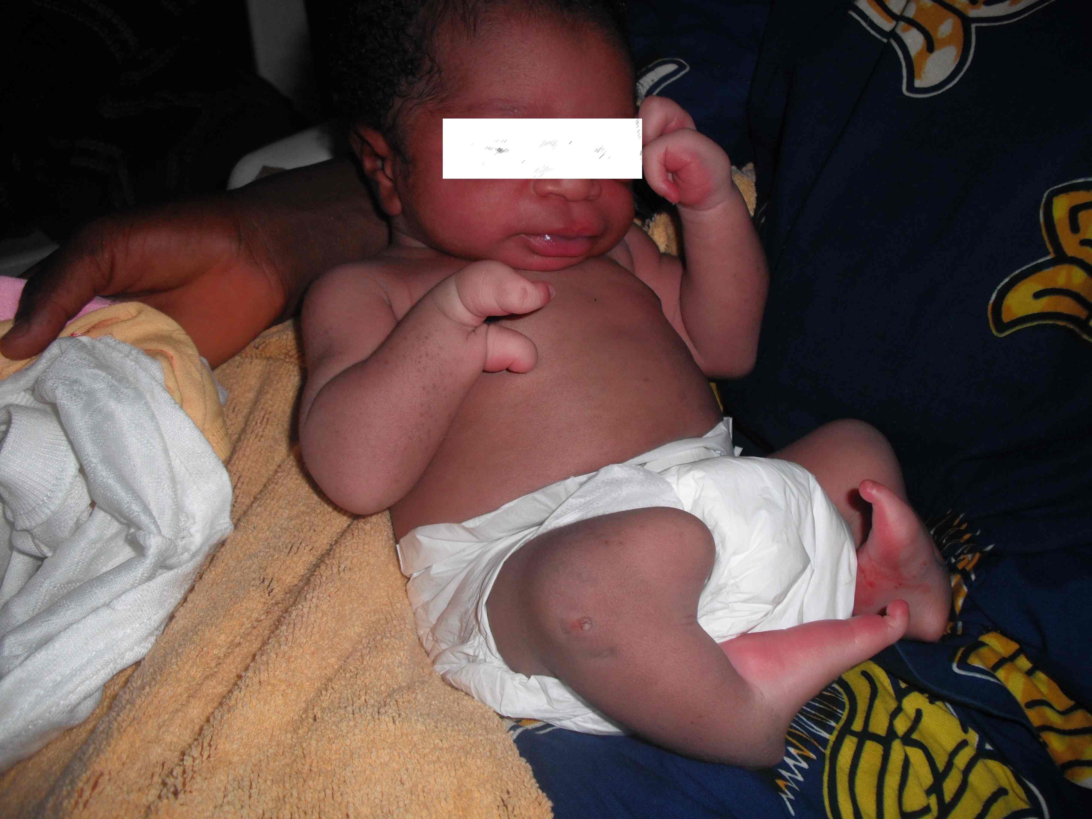
Figure 2: Both hands and legs.
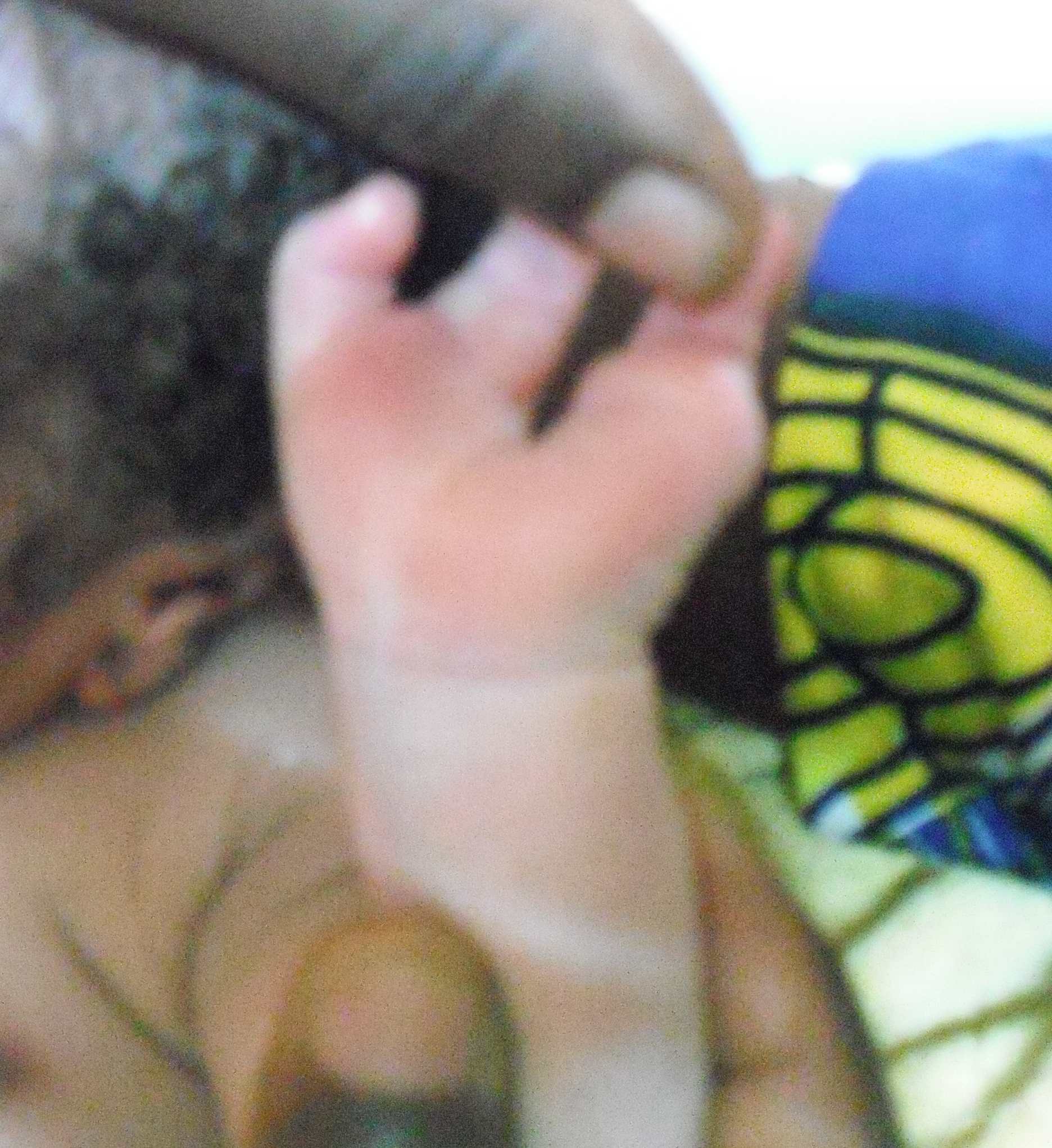
Figure 3: Clawed digits (Split hand).
The anterior fontanel was normotensive, about 4 cm x 6 cm. The baby had low set ears, no facial cleft or ocular abnormality. Anthropometry parameters were normal (weight 3.0 kg, OFC 36.0 cm, Length 49.5 cm).
Both arms and forearms were normal. The right upper limb had two digits with proximal and distal interphalangeal joints, but no carpal and no metacarpal bones. Ectrodactyly (cleft) of the left hand, with three digits (1st, 2nd, and 5th digits). The 1st digit attached more distally to the 2nd (Figs. 1-3). Bilateral hypoplastic legs with malformed flexed and internally rotated knee and ankle joints. Each foot had only a digit.
Ultrasound revealed no structural abnormality of intra-abdominal organs while babygram, (Figs. 4,5) showed hypoplasia of both tibia and fibula and dilated loops of the bowels. There was no spinal abnormality and other systems were normal. But the father had similar clefts of the left hand and the right foot, (Figs. 6-8). A diagnosis of familial ectrodactyly was made.
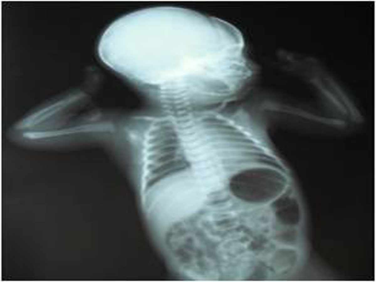
Figure 4: Babygram 1 showing dilated loops of bowels.
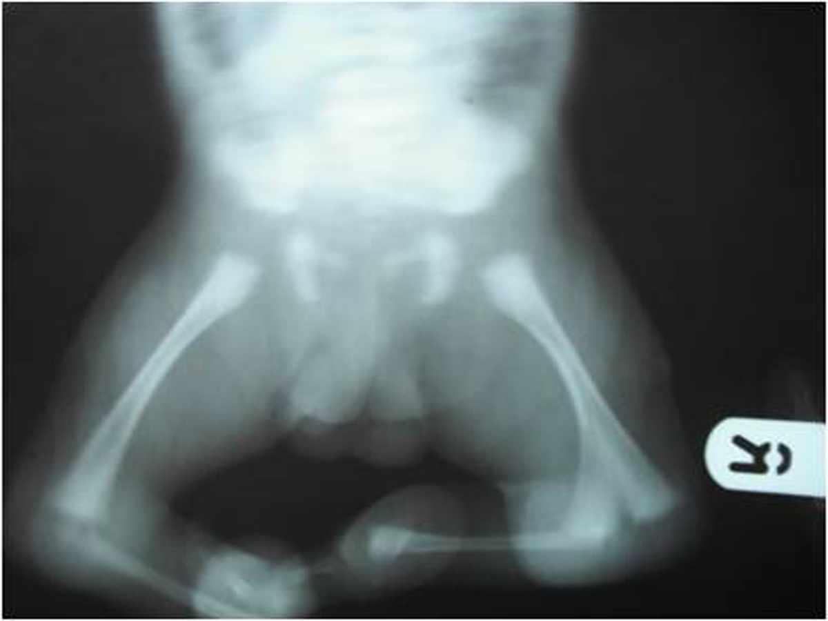
Figure 5: Babygram 2 showing hypoplasia of both tibia and fibula.
The parents were counseled and the child was discharged on the third day of life for follow-up care following the parents' persistence. The baby however was not presented for follow-up care, and was said to have died when a follow-up call was made to the parents two week after birth.
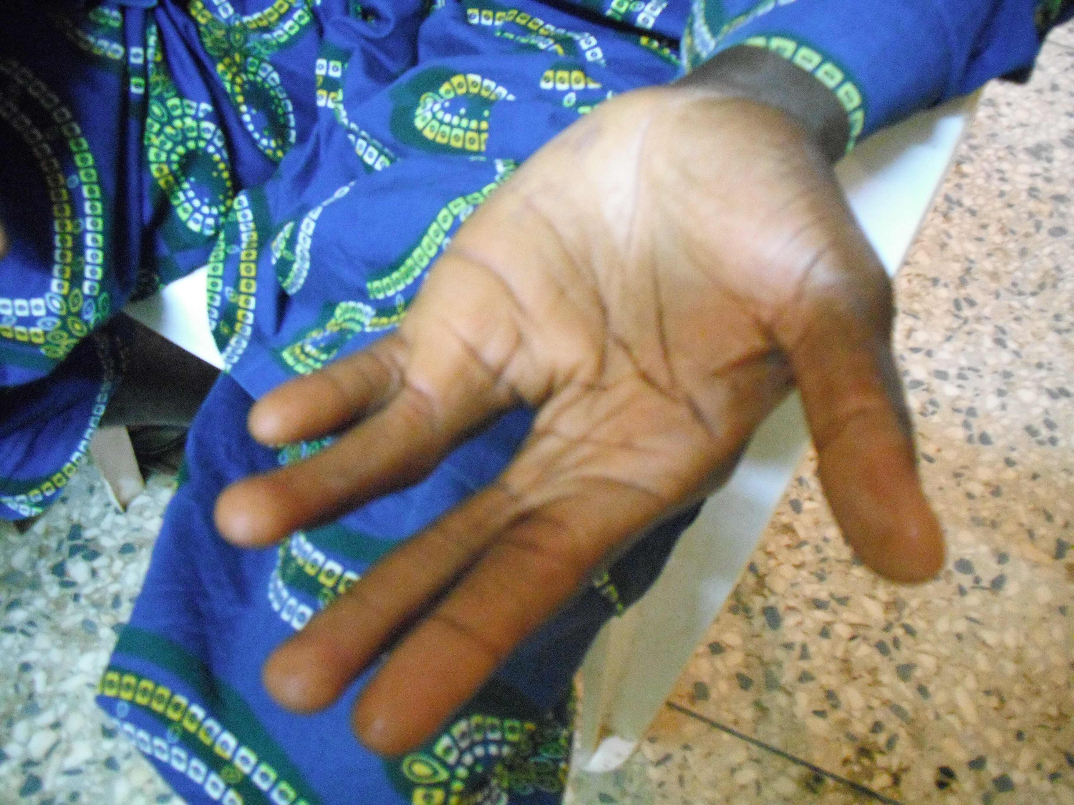
Figure 6: Father’s left hand
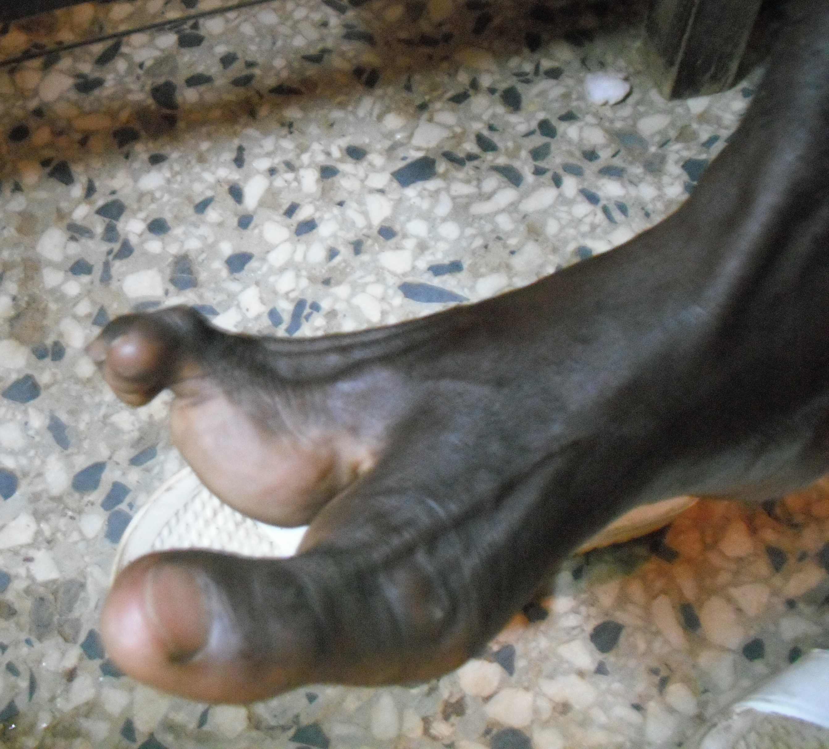
Figure 7: Father’s right foot.
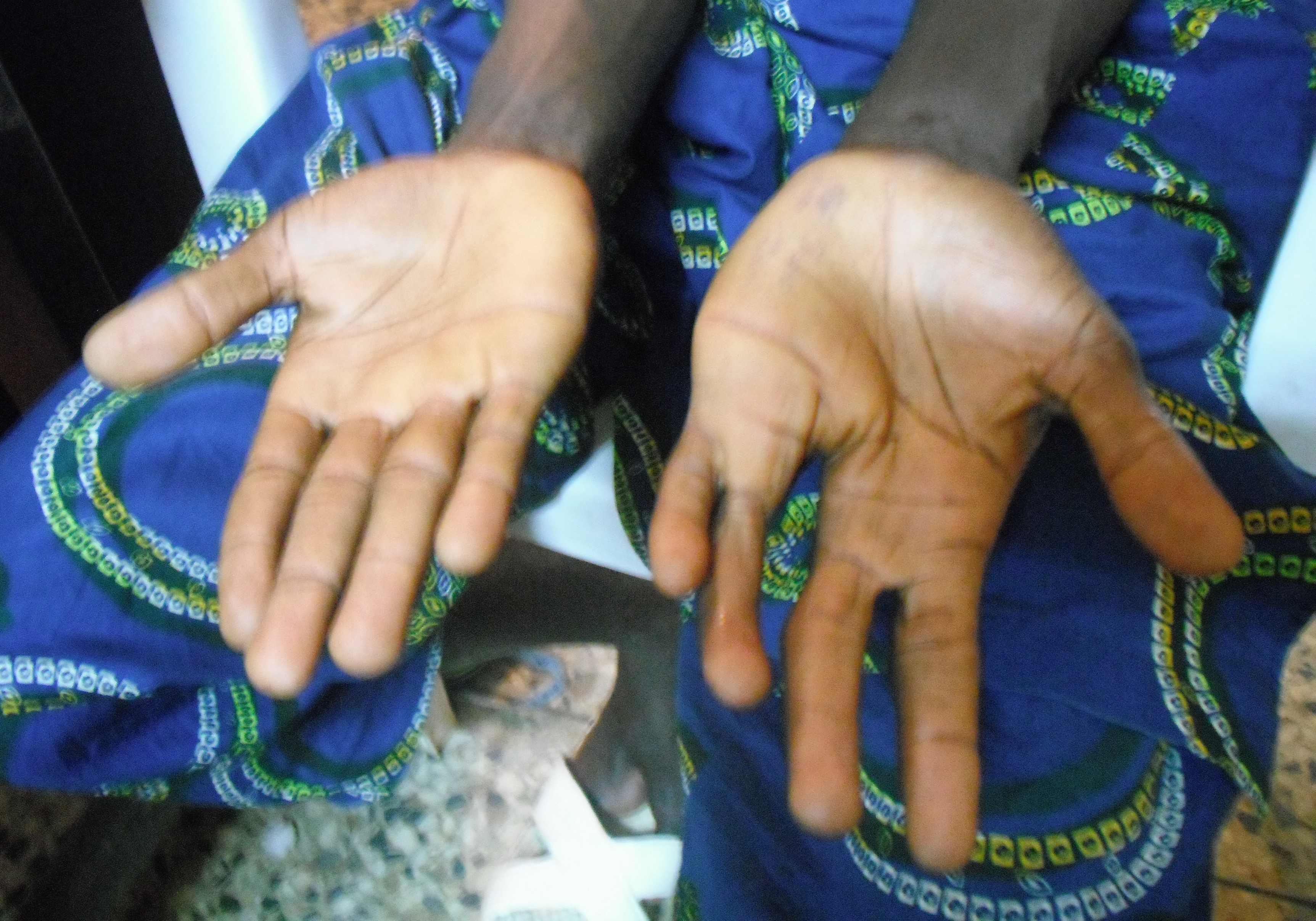
Figure 8: Father’s both hands.
Discussion
This is a case of familial ectrodactyly most likely with autosomal pattern of inheritance. The penetrance is obviously high in this case. Possible parental denial made it difficult to ascertain the involvement of the two siblings who reportedly had no malformation but died of febrile illness before their second birthdays. A patient with isolated ectrodactyly can live near normal life if well managed, just as the father of this infant who is a driver, but superstitious beliefs and social stigma constitute challenges to survival of such patients. Despite counseling, this child was not brought for follow up, and unfortunately, the managing unit was only told of his sudden death at two weeks of life when the team made a follow-up call. The outcome of this infant is not different from many cases of major congenital malformation in developing countries, where parents’ initial enthusiasm to seek medical help become dashed when they soon realized that the much anticipated cure may not exist.11 Some parents and relatives readily think infanticide may be a way of coping with the associated social stigma. Illiteracy and declining primary healthcare negatively impact the health seeking behavior of parents, this partly explain the reason this woman with two infantile death and a family risk factor for congenital malformation still did not benefit from prenatal diagnosis, counseling and management. Facilities for chromosomal studies are not readily available in most developing countries like Nigeria, this limits understanding of the genetic disorder underlining most of the congenital abnormalities encountered in clinical practice. Physicians need to rise up to the challenges in management of children with congenital malformations such as ectrodactyly, and be equipped to answer without unnecessary delay the usually burning questions in the minds of the parents, namely; what is this? what is the cause? Does it have any cure? and is there a chance of recurrence? The quality of counseling, social support and expertise for the required multidisciplinary care will determine if we shall make progress in achieving improved survival and quality of life of children with major congenital malformation.
Major congenital malformations contribute to infant morbidity and mortality, the prevalence in developing countries is largely unknown, and current preventive public health approach may not significantly impact the incidence. Acquisition of management skills especially quality counseling, availability of diagnostic tools, especially chromosomal studies and multidisciplinary care will contribute significantly to improve survival of a child with congenital malformations such as ectrodactyly.
Conclusion
In summary, there is need for clinicians to be better prepared for management of all aspects, including the social aspects of children with major congenital anomalies. Counseling of the parents and the care givers should be provided irrespective of the similarity of the anomaly with that seen on the parent(s).
Acknowledgements
The authors reported no conflict and no funding was received on this work.
|