The term ovarian cancer is used to describe any malignancy that involves the ovaries.1 It is the sixth major cause of cancer fatalities in women, ranking seventh among all female cancers, and accounting for approximately 4% of all incidences of cancer. Globally, approximately 225 000 new cases are diagnosed each year, with nearly 140 000 deaths. Though the overall prevalence of ovarian cancer is higher in industrialized countries, age-specific mortality rates tend to be higher in the developing world.2 A substantial and rapidly rising proportion of the world’s cancer patients is in Asia, home of about 60% of the world’s population.3 There are several reasons for this trend. First, the rapid increases in public awareness and availability of cancer screening facilities have significantly increased the number of diagnosed cases in Asia.4 Second, lifestyle changes such as sedentary behavior, unhealthy diet, and increased consumption of alcohol and tobacco. A third major contributor is the aging of populations across Asian nations, which has elevated the risk for most cancers due to accumulated genetic damage and weakened cellular repair.5
The escalating cancer burden worldwide has spurred intense research efforts aimed at improving early detection, prognostication, and treatment outcomes. Among the prognostic markers widely studied in ovarian cancer are the platelet-to-lymphocyte ratio (PLR) and neutrophil-to-lymphocyte ratio (NLR).
The PLR is a repeatable, affordable, and widely available hematological marker. In patients with malignancies, PLR has been used as an indicator of systemic inflammation and thrombotic tendency, which may explain its potential prognostic value.6–8 Elevated PLR is associated with the activation of key transcription factors involved in inflammation, including nuclear factor-kB, signal transducer and activator of transcription 3, and hypoxia-inducible factor-1 alpha in the inflammatory response. These, in turn, trigger the production of tumor-promoting cytokines such as tumor necrosis factor-alpha, interleukin-1, and interleukin-6.9
Additionally, NLR has emerged as one of the most widely used hematological markers.10 It has consistently been shown to predict survival outcomes not only in cancer patients, but also in individuals with chronic inflammatory, cardiovascular, infectious, and immune-related conditions, as well as in the general population.11 Across all cancer types, elevated NLR levels above established thresholds have consistently been associated with poorer outcomes.12 However, in ovarian cancer, where diverse subtypes and classifications can influence prognosis, the use of NLR and PLR has been limited to cases with general malignancy, leading to uncertainty in their predictive power, which requires comprehensive evaluation. Therefore, this study aimed to evaluate the role of NLR and PLR as prognostic biomarkers for ovarian cancer Asian populations through a meta-analysis approach.
Methods
This systematic review and meta-analysis assessed the association of ovarian cancer with PLR and NLR as prognostic markers in Asian populations based on the Preferred Reporting Items for Systematic Reviews and Meta-Analysis protocol.13 Being based on data from previous studies, ethical approval was not required.
Identification of relevant literature
A systematic search was conducted in online databases, including PubMed, Cochrane Library, ScienceDirect, and Google Scholar, to identify relevant studies. The following Boolean search string was used: (“PLR” OR “Platelet-to-Lymphocyte Ratio” OR “NLR” OR “Neutrophil-to-Lymphocyte Ratio”) AND (“Ovarian Cancer”) AND (“Prognosis”). All authors participated in the initial screening process, followed by an independent evaluation of each study based on predetermined eligibility criteria. The final selection of studies to include was determined through group discussion are required unanimous agreement. Any disagreements among reviewers were resolved by consensus.
Eligibility criteria
Five independent investigators carefully screened the candidate publications. Studies were considered eligible if they fulfilled the following criteria: (1) Original research articles which included patients diagnosed with ovarian cancer; (2) Studies that included the inflammatory markers NLR and/or PLR with cut-off values; and (3) Studies reporting the prognostic outcomes based on NLR and/or PLR evaluation, with sufficient data of hazard ratio (HR) and 95%CI for disease-free survival (DFS) or event-free survival (EFS) or progression-free survival (PFS), or overall survival (OS).
Data extraction
The following correlative information was collected in a predefined table: general study information (first author, publication year, and country), patient characteristics including treatment status, type of cancer, number of patients with types of cancer with poor prognosis (including serous, clear-cell, and mucinous), number with advanced-stage disease (III and IV), disease grades, presence of infection, and use of nonsteroidal anti-inflammatory drugs (NSAIDs) and corticosteroids, as well as endpoint parameters (DFS/EFS/PFS and OS).
Assessment of risk of bias
The Cochrane Risk of Bias 2 (RoB2) tool was used to evaluate the methodological and reporting biases in the included studies. Two impartial reviewers assessed the risk of bias and assigned ratings in each category: ‘low bias,’ ‘moderate concerns,’ or ‘high bias.’
Statistical analysis
The primary outcomes in this study were survival outcomes, which included DFS/EFS/PFS and OS presented as HR. The heterogeneity of the studies was determined using the Cochran Q-test and I2 statistic on a random effects model, where I2 > 50% and p < 0.10 were considered indicators of apparent heterogeneity. The sources of heterogeneity were determined using subgroup and sensitivity analyses. Funnel plots with Egger’s tests were adopted for more precise assessment of potential publication bias. Statistical analyses were performed using Review Manager (RevMan), version 5.4.1 (The Cochrane Collaboration, London, UK); A p-valu < 0.05 was considered statistically significant.
Results
Study Selection
The initial search yielded 337 articles. After removing the duplicates using Citation Manager software, 303 articles remained. After screening titles and abstracts, 57 articles were identified for evaluation, of which 39 were excluded because they could not be retrieved. The remaining 20 studies, which satisfied the selection criteria, were included. The study selection process is illustrated in the The Preferred Reporting Items for Systematic Reviews and Meta-Analysis flowchart [Figure 1].
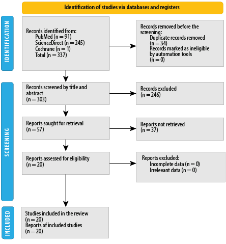 Figure 1: The Preferred Reporting Items for Systematic Reviews and Meta-Analysis Flowchart of the selection process.
Characteristics of Reviewed Studies
A total of 20 studies comprising 5316 ovarian cancer patients met the inclusion criteria and were selected for this review. There were nine studies from China (N = 3328, 62.8%), five from South Korea (1358, 25.5%), two from Japan (427, 8.0%), two from Thailand (92, 1.7%), and one from Israel (111, 2.1%). Thus, the overall patient ethnicity was 96.2% East Asian. Detailed characteristics of the studies are presented in Table 1. Egger’s test showed no potential publication bias. No information regarding the use of NSAIDs or corticosteroids was found in any of the studies.
Table 1: Characteristics of selected 20 studies evaluating epithelial ovarian cancer (EOC) cases in Asian populations (N = 5316).
|
Chen et al,14 2020, China
|
52.0
|
Post-treatment (surgery)
|
84
|
Clear-cell 100%
|
Clear-cell 100%, 84 (100)
|
37 (44.0)
|
I–II 47 (56.0);
III–IV 37 (44.0)
|
N/R
|
Yes
|
|
Chon et al,15 2020,
S. Korea
|
N/R
|
Post-treatment (surgery + platinum-based chemo)
|
102
|
Serous 73.5%, mucinous 3.9%, clear-cell 6.9%
|
Serous 73.5%, mucinous 3.9%, clear-cell 6.9%, 86 (84.3)
|
102 (100)
|
III 83 (81.4);
IV 19 (18.6)
|
G1 5 (4.9),
G2 37 (36.3),
G3 60 (58.8)
|
Yes
|
|
Eo et al,16 2016,
S. Korea
|
54.0
|
Post-treatment (surgery)
|
234
|
Serous 56.4%, mucinous 15.0%, clear-cell 15.0%
|
202 (86.3)
|
137 (58.5)
|
I–II 97 (41.5);
III–IV 137 (58.5)
|
G1 46 (19.7),
G2 65 (27.8),
G3 113 (48.3),
N/R 10 (4.3)
|
Yes
|
|
Feng et al,17 2016, China
|
56.0
|
Post-treatment (surgery)
|
875
|
High-grade serous 100%
|
875 (100)
|
800 (91.4)
|
I–II 75 (8.6);
III–IV 800 (91.4)
|
N/R
|
N/R
|
|
Kwon et al,18 2017
S. Korea
|
70.0
|
Post-treatment (surgery + platinum-based chemo)
|
42
|
Serous 78.6%
|
33 (78.6)
|
42 (100)
|
III 35 (83.3);
IV 7 (16.7)
|
G1 7 (16.7),
G2/3 35 (83.3)
|
N/R
|
|
Kwon et al,19 2018,
(S. Korea)
|
50.0
|
Post-treatment (surgery + platinum-based chemo)
|
109
|
Clear-cell 100%
|
109 (100)
|
45 (41.3)
|
I–II 64 (58.7);
III–IV 45 (41.3)
|
N/R
|
N/R
|
|
Miao et al,20 2016, China
|
55.0
|
Post-treatment (surgery + platinum-based chemo)
|
344
|
Serous 66.8%
|
216 (62.8)
|
176 (51.2)
|
I–II 168 (48.8); III–IV 176 (51.2)
|
G1/2 136 (39.5), G3 208 (60.5)
|
N/R
|
|
Paik et al,8 2016,
S. Korea
|
51.0
|
Post-treatment (surgery + adjuvant chemo)
|
674
|
Serous 63.4%, mucinous 8.6%, clear-cell 8.2%
|
540 (80.1)
|
451 (66.9)
|
I 150 (22.3),
II 73 (10.8),
III 389 (57.7),
IV 62 (9.2)
|
G1 55 (8.2),
G2 146 (21.7),
G3 473 (70.2)
|
N/R
|
|
Salman et al,21 2018, Israel
|
61.8 and 67.0
|
Post-treatment (surgery + adjuvant chemo)
|
111
|
Serous 59.5%, mucinous + clear-cell 10.8%
|
78 (70.3)
|
111 (100)
|
IIIC 88 (79.3);
IV 23 (20.7)
|
N/R
|
N/R
|
|
Supoken et al,22 2014, Thailand
|
52.0
|
Post-treatment (adjuvant paclitaxel and carboplatin)
|
36
|
Clear-cell 100%
|
36 (100)
|
16 (44.4)
|
I 17 (47.2),
II 3 (8.3),
III 11 (30.6),
IV 5 (13.9)
|
N/R
|
N/R
|
|
Wang et al,23 2016, China
|
52.3
|
Post-treatment (surgery)
|
143
|
85% unspecified
|
122 (85.3)
|
89 (62.2)
|
I–II 54 (37.8);
III–IV 89 (62.2)
|
N/R
|
Yes
|
|
Tang et al, 2020, China24
|
50.0
|
Post-treatment (adjuvant chemo)
|
214
|
Serous 69.3%, mucinous 4.9%, clear-cell 5.3%
|
179 (83.6)
|
124 (58.0)
|
I 62 (27.6),
II 39 (17.3),
III 109 (48.4),
IV 15 (7.0)
|
G1 42 (19.6), G2 48 (22.4), G3 135 (63.1)
|
N/R
|
|
Yoshida et al,25 2019,
Japan
|
53.0
|
Post-treatment (surgery)
|
83
|
Clear-cell 100%
|
83 (100)
|
0 (0.0)
|
I 76 (91.6),
II 7 (8.4)
|
N/R
|
N/R
|
|
Zhang et al,26 2015, China
|
50.6
|
Post-treatment (surgery + platinum-based chemo)
|
190
|
Serous 53.2%, mucinous 6.3%, clear-cell 3.7%
|
120 (63.2)
|
137 (72.1)
|
I 22 (11.6),
II 31 (16.3),
III 128 (67.4),
IV 9 (4.7)
|
G1 64 (33.7),
G2 44 (23.2),
G3 69 (36.3)
|
Yes
|
|
Zhou et al,27 2018, China
|
54.3
|
Post-treatment (surgery + platinum-based chemo)
|
370
|
Serous 64.3%, mucinous 0.8%, clear-cell 1.1%
|
245 (66.2)
|
370 (100)
|
III 370 (100)
|
G1 24 (6.5),
G2 133 (35.9),
G3 213 (57.6)
|
N/R
|
|
Li et al,28 2020, China
|
N/R
|
Pre-treatment
|
117
|
Serous 73.5%
|
86 (73.5)
|
72 (61.5)
|
I–II 45 (38.5),
III–IV 72 (61.5)
|
G1 67 (57.3),
G2–G3 50 (42.7)
|
Yes
|
|
Kim et al,29 2018,
S. Korea
|
57.0
|
Pre-treatment
|
197
|
High-grade serous 91.4%
|
180 (91.4)
|
197 (100)
|
III 52 (26.4),
IV 145 (73.6)
|
N/R
|
N/R
|
|
Komura et al,30 2017,
Japan
|
N/R
|
Post-treatment (surgery)
|
344
|
Serous 38.1%, mucinous 9.3%, clear-cell 24.1%
|
246 (71.5)
|
155 (45.1)
|
I–II 189 (54.9), III–IV 155 (45.1)
|
N/R
|
N/R
|
|
Sowannakul et al,31 2023,
Thailand
|
54.5
|
Post-treatment (platinum-based chemo)
|
56
|
Serous 55.4%, mucinous 5.4%, clear-cell 21.4%
|
46 (82.1)
|
43 (76.8)
|
I 11 (19.6),
II 2 (3.6),
III 29 (51.8),
IV 14 (25.0)
|
N/R
|
Yes
|
N/R: Not reported; G1–G3: Tumor grades 1 to 3;Chemo: Chemotherapy.
Neutrophil-to-Lymphocyte Ratio
Univariate analysis of NLR showed a significant relationship between NLR and OS in the overall effect analysis with a random effect model showing HR = 2.82 (95% CI: 1.63–4.88; p < 0.001; overall I2 = 97.1%). In the subgroup analysis, various patient groups showed significant results. For patients who underwent only surgery post-treatment random effect model revealed HR = 3.13 (95% CI: 2.17–4.50; I2 = 69.9%). For the group of patients who received a combination of surgery and chemotherapy random effect model showing HR = 2.62 (95% CI: 1.03–6.6; I2 = 97.9%), the group who received only chemotherapy contained one study by Sowannakul et al,31 with HR = 2.72 (95% CI: 1.31–5.63). There was no significant difference in subgroup analysis with the random effects model (χ2 = 0.20, p = 0.904) [Figure 2].
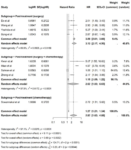 Figure 2: Overall survival based on univariate analysis of neutrophil-to-lymphocyte ratio between the subgroups who underwent surgery alone,16,23,25,32 surgery with chemotherapy,18,20,21,26 and post-chemotherapy only.31
Multivariate analysis of NLR showed a significant relationship between NLR and OS in the overall effect analysis with a random effect model showing HR = 1.44 (95% CI: 1.16–1.80; p < 0.001; overall I2 = 70.0%). In the subgroup analysis, significant results were found in the study group with patients with post-treatment status who only underwent surgery with a random effect model showing HR = 2.51 (95% CI: 0.89–7.10; I2 = 78.5%). The study group with patients who received a combination of surgery and chemotherapy had significant results with a random effect model showing HR = 1.30 (95% CI: 1.01–1.67; I2 = 70.5%). However, the study group with patients who had not received treatment (pre-treatment) had only one study by Kim et al,29 with HR = 1.89 (95% CI: 1.11–3.24), and the study group with patients who only received chemotherapy had one study by Sowannakul et al,31 with HR=1.25 (95% CI: 0.40–3.92). There was no significant difference in subgroup analysis with a random effects model (χ2 = 2.76, p = 0.430) [Figure 3].
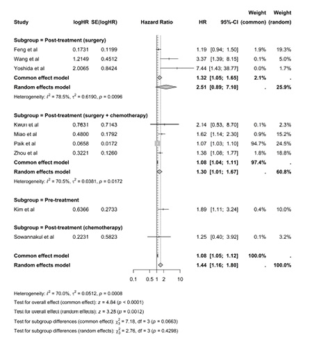 Figure 3: Overall survival based on multivariate analysis of neutrophil-to-lymphocyte ratio among the subgroups who underwent surgery alone,17,23,25 surgery with chemotherapy,8,18,20,27 pre-treatment status,29 and post-chemotherapy only.31
Univariate analysis of NLR showed a significant relationship between NLR and DFS/EFS/PFS in the overall effect analysis with a random effect model showing HR = 2.43 (95% CI: 1.59–3.73; p <0.001; overall I2 = 91.2%). In the subgroup analysis, significant results were found in the study group with patients with post-treatment status who only underwent surgery with a random effect model showing HR = 1.99 (95% CI: 1.28–3.10; I2 = 84.2%). The study group with patients who received a combination of surgery and chemotherapy had significant results with a random effect model showing HR = 3.28 (95% CI: 1.60–6.71; I2 = 89.8%), and the study group with patients who only received chemotherapy only contained one study by Sowannakul et al,31 with HR = 2.57 (95%CI: 1.37–4.81). There was no significant difference in subgroup analysis with the random effects model (χ2 = 1.46, p = 0.482) [Figure 4].
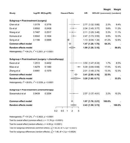 Figure 4: Disease-free survival/event-free survival/progression-free survival based on univariate analysis of neutrophil-to-lymphocyte ratio among the subgroups who underwent surgery alone,14,16,23,30,32 surgery with chemotherapy,18,20,26 and post-chemotherapy only.31
Multivariate analysis of NLR showed a significant relationship between NLR and DFS/EFS/PFS in the overall effect analysis with a random effect model showing HR = 1.41 (95% CI: 1.17–1.69; p < 0.001; overall I2 = 69.3%). In the subgroup analysis, no significant results were found in the study group with patients with post-treatment status who only underwent surgery with a common effect model showing HR = 1.32 (95% CI: 1.13–1.55; I2 = 30.8%). The group with patients who received a combination of surgery and chemotherapy had significant results with a random effect model showing HR = 1.36 (95% CI: 0.95–1.93; I2 = 69.5%), the group with patients who only received chemotherapy only contained one study by Sowannakul et al. with HR = 1.52 (95% CI: 0.54–4.31), the pre-treatment group showed a significant result with HR = 1.53 (95% CI: 1.02–2.28; I2 = 53.0%).31 There was no significant difference in subgroup analysis with the random effects model (χ2 = 0.21, p = 0.976) [Figure 5].
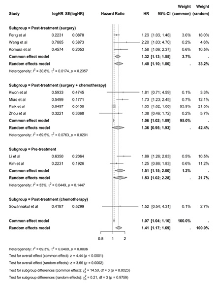 Figure 5: Disease-free survival, event-free survival or progression-free survival based on multivariate analysis of neutrophil-to-lymphocyte ratio among the subgroups who underwent surgery alone,17,23,30 surgery with chemotherapy,8,18,20,27 pre-treatment status,28,29 and post-chemotherapy.31
Platelet-to-Lymphocyte Ratio
Univariate analysis of PLR showed a significant relationship between PLR and OS in the overall effect analysis with a random effect model showing HR = 2.00 (95% CI: 1.37–2.92; p <0.01; overall I2 = 92.0%). In the subgroup analysis, significant results were found in the study group with patients with post-treatment status who only underwent surgery with a random effect model showing HR = 1.72 (95% CI: 1.06–2.79; I2 = 75.0%). The study group with patients who received a combination of surgery and chemotherapy had significant results with a random effect model showing HR = 2.13 (95% CI: 1.09-4.18; I2 = 96.0%), and the study group with patients who only received chemotherapy only contained one study by Sowannakul et al.31 with HR = 2.20 (95% CI: 1.02–4.77). There was no significant difference in subgroup analysis with the random effects model (χ2 = 0.41, p = 0.815) [Figure 6].
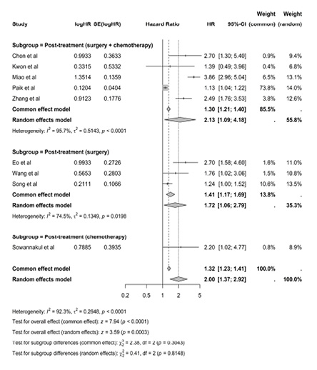 Figure 6: Overall survival based on univariate analysis of platelet-to-lymphocyte ratio among subgroups who underwent surgery with chemotherapy,8,15,18,20,26 surgery only,16,23,32 and post-chemotherapy only.31
Multivariate analysis of PLR showed a significant relationship between PLR and OS in the overall effect analysis with a random effect model showing HR = 1.82 (95% CI: 1.31–2.54; p = 0.008; overall I2 = 68.2%). In the subgroup analysis, the study group with patients with post-treatment status who only underwent surgery with a common effect model did not show significant results with HR = 1.20 (95% CI: 0.97–1.47; I2 = 34.4%), the study group with patients who received a combination of surgery and chemotherapy had significant results with a common effect model showing HR = 2.13 (95% CI: 1.69–2.68; I2 = 0.0%), and the study group with patients who only received chemotherapy only contained one study by Sowannakul et al,31 with HR = 2.54 (95% CI: 0.93–6.39).There was no significant difference in subgroup analysis with the random effects model (χ2 = 3.81, p = 0.149) [Figure 7].
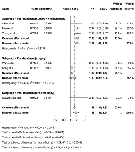 Figure 7: Overall survival based on multivariate analysis of platelet-to-lymphocyte ratio between the subgroups who underwent surgery with chemotherapy,15,20,26 surgery alone,23,32 and post-chemotherapy only.31
Univariate analysis of PLR showed a significant relationship between PLR and DFS/EFS/PFS in the overall effect analysis with a random effect model showing HR = 1.85 (95% CI: 1.32–2.59; p <0.001; overall I2 = 92.1%). In the subgroup analysis, the study group with patients who only underwent surgery with a common effect model did not show significant results with HR = 1.32 (95% CI: 1.13–1.54; I2 = 44.1%), the study group with patients who received a combination of surgery and chemotherapy had significant results with a random effect model showing HR = 2.09 (95% CI: 1.01–4.33; I2 = 96.7%), and the study group with patients who only received chemotherapy with a random effect model did not show significant results with HR = 2.22 (95% CI: 0.73-6.73; I2 = 69.1%). There was no significant difference in subgroup analysis with the common effects model (χ2 = 2.31, p = 0.316) [Figure 8].
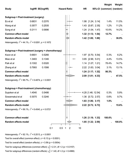 Figure 8: Disease-free survival/event-free survival/progression-free survival based on univariate analysis of platelet-to-lymphocyte ratio among the subgroups who underwent surgery alone,16,23,32 surgery with chemotherapy,8,18,20,26 and post-chemotherapy only.22,31
Multivariate analysis on PLR showed a significant relationship between PLR and DFS/EFS/PFS in the overall effect analysis with a random effect model showing HR = 1.47 (95% CI: 1.02-2.12; p < 0.001; overall I2 = 76.5%). In the subgroup analysis, the study group with patients who only underwent surgery with a random effect model did not show significant results with HR = 1.11 (95% CI: 0.61-2.03; I2 = 61.9%). The study group with patients who received a combination of surgery and chemotherapy had significant results with a common effect model showing HR = 1.80 (95% CI: 1.45–2.23; I2 = 0.0%), but the study group with patients who only received chemotherapy with a random effect model did not show significant results with HR = 1.81 (95% CI: 0.38–8.62; I2 = 67.8%). There was no significant difference in subgroup analysis with the random effects model (χ2 = 2.18, p = 0.336) [Figure 9].
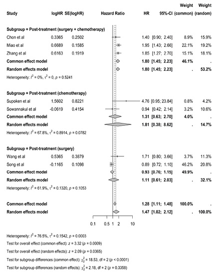 Figure 9: Disease-free survival/event-free survival/progression-free survival based on multivariate analysis of platelet-to-lymphocyte ratio among the subgroups who underwent surgery with chemotherapy,15,20,26 chemotherapy only,22,31 and surgery only.23,32
Discussion
This meta-analysis of 20 studies evaluated the prognostic value of systemic inflammatory markers, NLR and PLR, in Asian ovarian cancer patients. This study found that high levels of NLR and PLR are consistently associated with poorer OS and prognostic outcomes. These were confirmed by univariate and multivariate analyses, establishing their independent prognostic value for clinicians.
Neutrophil-to-Lymphocyte Ratio
Both univariate and multivariate analyses showed a significant negative association between NLR and OS and PFS. This aligns with the recent meta-analysis by Zhang et al,23 who also reported a strong inverse correlation between NLR and PFS with OR (HR ≈ 1.21; p < 0.001). The mechanism underlying the association between high NLR and poor cancer prognosis remains unclear.33 It is possible that an increase in NLR may reflect both an increase in neutrophil-mediated inflammatory response and a decrease in lymphocyte-mediated anti-tumor immune response, facilitating tumor growth. This is supported by Milne et al,34 who found the lymphocyte counts of ovarian cancer patients to be significantly lower at diagnosis compared to two years prior, with a greater rate of decline at advanced stages. Thus, accelerated tumor growth is associated with poor survival outcomes.
The observed differences between univariate and multivariate analyses in our study appear to be due to variations in data availability in included studies. The high heterogeneity could be attributable to regional differences, sample sizes, patient characteristics, types of therapy, and NLR cut-off values.
Our subgroup analyses showed a significant association between OS and PFS with NLR based on pre-treatment and post-treatment settings, including surgery, chemotherapy, or both. While Wang et al,23 suggested pre-treatment NLR to be a viable predictor for malignancy and advanced tumor stage, Topcu et al,19 viewed it as an ineffective marker. In ovarian clear cell carcinoma, preoperative NLR level is associated with postoperative prognostic indicators, such as International Federation of Gynecology and Obstetrics stage, residual tumor, and platinum-based chemotherapy resistance.35 Thus, post-treatment NLR levels can be used to identify high-risk patients.15 A study on 49 ovarian cancer patients found that higher NLR was significantly associated with shorter survival or poorer prognosis after chemotherapy.36 A meta-analysis of 10 studies with 2919 patients reported that patients with higher NLR pre-treatment levels had relatively shorter OS (HR = 2.36) and PFS (HR = 1.82).37 In addition, high NLR reflects the formation of an immunosuppressive tumor environment that encourages metastasis. Therefore, NLR level can also serve as a predictive marker for metastasis.38
Platelet-to-Lymphocyte Ratio
PLR was similarly associated with survival outcomes across the studies we analyzed. Well-aligned with our results is the meta-analysis by Tian et al,25 with a total of 11 studies comprising 3574 patients with ovarian cancer. The random-effects meta-analysis demonstrated that patients with elevated PLR had shorter OS (HR = 1.48, 95% CI: 1.24–1.76, p < 0.001) and PFS (HR = 1.38, 95% CI: 1.17–1.63, p < 0.001). The negative prognostic impact of high PLR on OS and PFS remained substantial in Asian populations, patients with PLR ≥ 200, and studies with Newcastle-Ottawa Scale score ≥ 7.39
Numerous studies suggest that the inflammatory response to tumor progression involves cross-talk between tumor cells, immune cells, and the tumor microenvironment.40 Platelets act as a reservoir for growth factors that regulate angiogenesis in cell proliferation, malignancies, metastasis, and migration.41 Lymphocytes infiltrate the cancer cells to induce cytotoxicity.42 Thus, a low lymphocyte count may compromise the immune response.43 This has been employed as a prognostic indicator in various cancers, including ovarian cancer.44
In this study, multivariate and univariate analyses of OS and PFS against PLR showed significant associations (p < 0.001) in each post-treatment subgroup of patients who underwent surgery, chemotherapy (neoadjuvant and/or adjuvant), or both surgery and chemotherapy. Increased platelet production, stimulated by megakaryocytes, raises the platelet count, which is correlated with an increased release of inflammatory mediators, supporting the link between elevated PLR and ovarian cancer recurrence.45 Additionally, recent research has demonstrated that neoadjuvant chemotherapy increases lymphocyte count, which is predictive of improved OS and PFS.46 This contradicts earlier research which associated neoadjuvant chemotherapy delivery with reduced platelet count, indicating worse prognosis.47
Ovarian cancer triggers an adaptive immune response due to its high mutation burden and the identification of the mechanisms underlying cancer mutations.48 The immune system helps restrict the proliferation of cancer cells by lymphocyte activity, improving patient outcomes.49 Kawahara et al,46 verified that the rise in tumor-infiltrating lymphocytes in peripheral blood flow prior to surgery had a suppressive effect on tumor cells and treatment sensitivity. These findings suggest that more aggressive treatments such as extended adjuvant chemotherapy may be required for better outcomes.
Rising levels of PLR and NLR reflect increasing inflammation within tumor cells. Inflammatory mediators, including cytokines and chemokines, are correlated with an increase in platelets and leukocytes.50 While immune responses help inhibit cancer growth, they can also enable inflammation-linked angiogenesis and immune evasion.51 Numerous factors, including platelets, lymphocytes, and neutrophils participate in these inflammatory processes. This may accelerate tumor growth in the direction of inflammation.52 Chronic inflammation supports carcinogenesis via several mechanisms, including proliferation, angiogenesis, invasion, metastasis, and the inhibition of cancer cell death.53 Research has demonstrated a connection between cancer cells and cytokines and chemokines. Cytokines are involved in controlling the growth and survival of cancer cells in ovarian cancer.51 Consequently, heightened inflammatory reactions, as shown by elevated PLR and NLR, might reliably predict the prognosis and response to treatment.54
In ovarian cancer, PLR > 300 is associated with a decline in OS and PFS, indicating worse prognosis.55 A meta-analysis involving 1250 patients with ovarian cancer reported a strong association between increased PLR and a decline in OS and PFS (HR = 1.63).56 Because these inflammatory indicators are accessible, affordable, and non-invasive when used in conjunction with laboratory data, their application in ovarian cancer is advantageous.50 Because of this, PLR is a better predictor of the incidence of inflammation in ovarian cancer than the neutrophil-linked indicators.20
Impact of Ethnicity
The studies included in our review were overwhelmingly East Asian (96.2%). In comparison, the two meta-analytical studies listed below have findings broadly consistent with ours regarding the prognostic value of PLR and NLR, but with higher pooled discriminatory values. Significant presence of other ethnicities may have influenced their outcomes.
- Ma et al,49 reviewed 12 studies on PLR, with the following ethnic composition: Caucasian (61.3%), East Asian (25.4%), and Southeast Asian (9.6%).56
- Zhao et al,57 reviewed 13 studies on NLR, with the following ethnic composition: East Asian (69.8%), Southeast Asian (8.5%), and Euro-American (21.7%) populations.
Indeed, previous studies on ovarian cancer outcomes have shown significant survival differences between different racial groups, with Asians exhibiting better outcomes. Prominent among these is a large SEER-based study by Fuh et al,52 which compared the ethnicities of 52 260 epithelial ovarian cancer patients in the United States. Asian-American patients had significantly higher five-year survival (59.1%) than Caucasian patients (47.3%); p < 0.001. At the same time, the fact that Asians presented at a younger age may also partly explain the better outcomes.
The increased mortality observed in low-income countries also raises concerns,53 given the disparity in socio-economic conditions in Asian countries.
NLR, PLR vs. Kinetic ELIMination
Though NLR and PLR show a good predictive value, they have lower discriminatory value compared to newer methods such as Kinetic ELIMination rate constant of CA-125 (KELIM). Corbaux et al,58 showed that patients with an unfavorable KELIM score had a 1.96-fold higher hazard of death than those with a favorable KELIM score (HR = 0.51 for OS). The PFS analysis also showed a higher discriminatory value for PFS (HR = 0.59). In addition, the prognostic value of KELIM remained stable through different time horizons, up to five years of follow-up.The stability of KELIM’s prognostic value, regardless of chemotherapy dosing schedules or the combination with additional drugs, such as bevacizumab or veliparib,59 suggests that KELIM reflects the intrinsic characteristics of cancer cells with greater discrimination.
However, NLR and PLR retain their utility.60 These markers can be easily determined from blood tests, and thus are simple, affordable, and easily accessible in everyday oncology practice, especially for many Asian patients unable to afford expensive tests such as KELIM.39
Additional Considerations
The strength of this study lies in the subgroup analysis conducted based on patient treatment status, which quantitatively states that there is no significant difference between treatment groups. Although, in theory, it can be explained that treatment status can affect the results of the study, the results of the analysis in this study showed the opposite, which are likely caused by other factors.
In several similar studies evaluating PLR and NLR in ovarian cancer, the presence of comorbid infection can be a significant factor and bias the results. Infection can increase the number of neutrophils and platelets and affect the ratio.61 Thus, it is argued that higher NLR and PLR values may not fully reflect the condition of ovarian cancer itself. Therefore, it is imperative to consider the patient’s infection status when analyzing these data. However, based on the articles included in the meta-analysis, most studies did not explicitly mention the infection status of the patients. This is a significant limitation as it is unclear whether the patient had an infection at the time of data collection, which may bias the results. Some studies stated that patients with infection at the time of data collection were excluded, but this was not consistent across the included articles, so the results obtained may have high variability.
In addition, the use of drugs such as NSAIDs, regular analgesics, and steroids can affect the results of NLR and PLR measurements. NSAIDs can affect the number of neutrophils, lymphocytes, and platelets in the body, which in turn may skew the NLR and PLR values. For example, NSAIDs can reduce the number of lymphocytes/neutrophils. At the same time, steroids can increase the number of neutrophil and platelet cells, which can lead to changes in the NLR and PLR ratios that do not fully reflect the cancer condition itself.62,63However, none of the included studies explicitly mentioned the use of these drugs. This is an important limitation, capable of generating bias.
The strength of this study lies in the subgroup analysis conducted based on patient treatment status, which quantitatively found no significant difference between the treatment groups. Although, in theory, treatment status can affect the results of the study, our analysis showed opposite results, which could potentially be due to certain unconsidered factors.
This study has several limitations. Although we aimed to represent Asian populations, which make up nearly 60% of humanity, no suitable studies from South Asia (~ 42% of the Asian population) could be found. The Middle East was also scarcely represented, with only a small study from Israel (2.1%), with no eligible studies from the Arab world or Iran. Southeast Asia contributed only one small study from Thailand (1.7%). Consequently, the study population was overwhelmingly East Asian (96.2%), limiting the generalizability of our findings largely to that region. At the same time, this imbalance highlights the need for high-quality research from South Asia, Central Asia, and the Middle East. There was potential for bias due to heterogeneity in the study populations. Differences in cancer stage, type, and grading could not be stratified clearly because the included studies could not be grouped and analyzed based on such variables. In addition, 82.7% of all included patients had ovarian cancer types with poor prognoses, such as serous, mucinous, and clear-cell types. Finally, there was a strong trend towards advanced-stage ovarian cancer patients with epithelial ovarian cancer subtypes of serous, mucinous, and clear-cell, but this could also be an indirect advantage of this study as it provided an overview of the prevailing trends.
Conclusion
Inflammatory markers such as PLR and NLR are satisfactory prognostic markers in ovarian cancer to predict patient outcome and survival. We found similar levels of inflammatory markers among patients who underwent surgery, chemotherapy, and both. However, these findings could be biased by the other variables not considered in this study. Therefore, more specific studies by strictly controlling the eligibility criteria are needed to validate and confirm these findings. In addition, we recommend more controlled studies from the unpresented regions such as South Asia, Central Asia, and the Middle East.
Disclosure
The authors declare no conflicts of interest. This study was self-funded by the authors and received no external funding.
Acknowledgments
We express our deepest gratitude to our faculty for supporting this study.
references
- Karlan BY, Alvarez RD, Strauss JF III. Evolving paradigms in research and care in ovarian cancers. Obstet Gynecol 2016 Oct;128(4):771-774.
- 2. Khazaei Z, Namayandeh SM, Beiranvand R, Naemi H, Bechashk SM, Goodarzi E. Worldwide incidence and mortality of ovarian cancer and Human Development Index (HDI): GLOBOCAN sources and methods 2018. J Prev Med Hyg 2021 Apr;62(1):E174-E184.
- 3. Ginsburg O, Bray F, Coleman MP, Vanderpuye V, Eniu A, Kotha SR, et al. The global burden of women’s cancers: a grand challenge in global health. Lancet 2017 Feb;389(10071):847-860.
- 4. Arnold M, Sierra MS, Laversanne M, Soerjomataram I, Jemal A, Bray F. Global patterns and trends in colorectal cancer incidence and mortality. Gut 2017 Apr;66(4):683-691.
- 5. Huang J, Ngai CH, Deng Y, Tin MS, Lok V, Zhang L, et al. Cancer incidence and mortality in Asian countries: a trend analysis. Cancer Control 2022;29:10732748221095955.
- 6. Xu H, He A, Liu A, Tong W, Cao D. Evaluation of the prognostic role of platelet-lymphocyte ratio in cancer patients treated with immune checkpoint inhibitors: A systematic review and meta-analysis. Int Immunopharmacol 2019 Dec;77:105957.
- 7. Krenn-Pilko S, Langsenlehner U, Thurner EM, Stojakovic T, Pichler M, Gerger A, et al. The elevated preoperative platelet-to-lymphocyte ratio predicts poor prognosis in breast cancer patients. Br J Cancer 2014 May;110(10):2524-2530.
- 8. Paik ES, Shim M, Choi HJ, Lee YY, Kim TJ, Choi CH, et al. Preoperative multiplication of neutrophil and monocyte counts as a prognostic factor in epithelial ovarian cancer. Cancer Biomark 2016;17(4):419-425.
- 9. Li B, Zhou P, Liu Y, Wei H, Yang X, Chen T, et al. Platelet-to-lymphocyte ratio in advanced Cancer: review and meta-analysis. Clin Chim Acta 2018 Aug;483:48-56.
- 10. Zou P, Yang E, Li Z. Neutrophil-to-lymphocyte ratio is an independent predictor for survival outcomes in cervical cancer: a systematic review and meta-analysis. Sci Rep 2020 Dec;10(1):21917.
- 11. Howard R, Kanetsky PA, Egan KM. Exploring the prognostic value of the neutrophil-to-lymphocyte ratio in cancer. Sci Rep 2019 Dec;9(1):19673.
- 12. Bartlett EK, Flynn JR, Panageas KS, Ferraro RA, Sta Cruz JM, Postow MA, et al. High neutrophil-to-lymphocyte ratio (NLR) is associated with treatment failure and death in patients who have melanoma treated with PD-1 inhibitor monotherapy. Cancer 2020 Jan;126(1):76-85.
- 13. Page MJ, McKenzie JE, Bossuyt PM, Boutron I, Hoffmann TC, Mulrow CD, et al. The PRISMA 2020 statement: an updated guideline for reporting systematic reviews. PLoS Med 2021 Mar;18(3):e1003583.
- 14. Chen W, Zhong S, Shan B, Zhou S, Wu X, Yang H, et al. Serum D-dimer, albumin and systemic inflammatory response markers in ovarian clear cell carcinoma and their prognostic implications. J Ovarian Res 2020 Aug;13(1):89.
- 15. Chon S, Lee S, Jeong D, Lim S, Lee K, Shin J. Elevated platelet lymphocyte ratio is a poor prognostic factor in advanced epithelial ovarian cancer. J Gynecol Obstet Hum Reprod 2021 Jun;50(6):101849.
- 16. Eo WK, Chang HJ, Kwon SH, Koh SB, Kim YO, Ji YI, et al. The lymphocyte-monocyte ratio predicts patient survival and aggressiveness of ovarian cancer. J Cancer 2016 Jan;7(3):289-296.
- 17. Feng Z, Wen H, Bi R, Ju X, Chen X, Yang W, et al. Preoperative neutrophil-to-lymphocyte ratio as a predictive and prognostic factor for high-grade serous ovarian cancer. PLoS One 2016 May;11(5):e0156101.
- 18. Kwon BS, Lee HJ, Yang J, Song YJ, Suh DS, Lee DH, et al. Prognostic value of preoperative lymphocyte-monocyte ratio in elderly patients with advanced epithelial ovarian cancer. Obstet Gynecol Sci 2017 Nov;60(6):558-564.
- 19. Kwon BS, Jeong DH, Byun JM, Lee TH, Choi KU, Song YJ, et al. Prognostic value of preoperative lymphocyte-monocyte ratio in patients with ovarian clear cell carcinoma. J Cancer 2018 Mar;9(7):1127-1134.
- 20. Miao Y, Yan Q, Li S, Li B, Feng Y. Neutrophil to lymphocyte ratio and platelet to lymphocyte ratio are predictive of chemotherapeutic response and prognosis in epithelial ovarian cancer patients treated with platinum-based chemotherapy. Cancer Biomark 2016 Jun;17(1):33-40.
- 21. Salman L, Sabah G, Jakobson-Setton A, Raban O, Yeoshoua E, Eitan R. Neutrophil-to-lymphocyte ratio as a prognostic factor in advanced stage ovarian carcinoma treated with neoadjuvant chemotherapy. Int J Gynaecol Obstet 2020 Jan;148(1):102-106.
- 22. Supoken A, Kleebkaow P, Chumworathayi B, Luanratanakorn S, Kietpeerakool C. Elevated preoperative platelet to lymphocyte ratio associated with decreased survival of women with ovarian clear cell carcinoma. Asian Pac J Cancer Prev 2014;15(24):10831-10836.
- 23. Wang YQ, Jin C, Zheng HM, Zhou K, Shi BB, Zhang Q, et al. A novel prognostic inflammation score predicts outcomes in patients with ovarian cancer. Clin Chim Acta 2016 May;456:163-169.
- 24. Tang Y, Hu HQ, Tang FX, Lin D, Shen R, Deng L, et al. Combined Preoperative LMR and CA125 for prognostic assessment of ovarian cancer. J Cancer 2020 Mar;11(11):3165-3171.
- 25. Yoshida K, Yoshikawa N, Shirakawa A, Niimi K, Suzuki S, Kajiyama H, et al. Prognostic value of neutrophil-to-lymphocyte ratio in early-stage ovarian clear-cell carcinoma. J Gynecol Oncol 2019 Nov;30(6):e85.
- 26. Zhang WW, Liu KJ, Hu GL, Liang WJ. Preoperative platelet/lymphocyte ratio is a superior prognostic factor compared to other systemic inflammatory response markers in ovarian cancer patients. Tumour Biol 2015 Nov;36(11):8831-8837.
- 27. Zhou M, Li L, Wang X, Wang C, Wang D. Neutrophil-to-lymphocyte ratio and platelet count predict long-term outcome of stage IIIC epithelial ovarian cancer. Cell Physiol Biochem 2018;46(1):178-186.
- 28. Li Z, Yin H, Ren M, Shen Y. Prognostic significance of CA125 dynamic change for progression free survival in patients with epithelial ovarian carcinoma. Med Sci Monit 2020 Sep;26:e925051.
- 29. Kim YJ, Lee I, Chung YS, Nam E, Kim S, Kim SW, et al. Pretreatment neutrophil-to-lymphocyte ratio and its dynamic change during neoadjuvant chemotherapy as poor prognostic factors in advanced ovarian cancer. Obstet Gynecol Sci 2018 Mar;61(2):227-234.
- 30. Komura N, Mabuchi S, Yokoi E, Kozasa K, Kuroda H, Sasano T, et al. Comparison of clinical utility between neutrophil count and neutrophil-lymphocyte ratio in patients with ovarian cancer: a single institutional experience and a literature review. Int J Clin Oncol 2018 Feb;23(1):104-113.
- 31. Sowannakul A, Rodpenpear N, Ekbhum P, Tantitamit T. Prognostic value of the neutrophil-to-lymphocyte ratio, platelet-to-lymphocyte ratio, and platelet count for platinum-sensitive recurrent epithelial ovarian cancer. Asian Pac J Cancer Prev 2023 Nov;24(11):3765-3771.
- 32. Song Q, Xu SX, Wu JZ, Ling L, Wang S, Shu XH, et al. The preoperative platelet to neutrophil ratio and lymphocyte to monocyte ratio are superior prognostic indicators compared with other inflammatory biomarkers in ovarian cancer. Front Immunol 2023 Jun;14:1177403.
- 33. Zugazagoitia J, Guedes C, Ponce S, Ferrer I, Molina-Pinelo S, Paz-Ares L. Current challenges in cancer treatment. Clin Ther 2016 Jul;38(7):1551-1566.
- 34. Milne K, Alexander C, Webb JR, Sun W, Dillon K, Kalloger SE, et al. Absolute lymphocyte count is associated with survival in ovarian cancer independent of tumor-infiltrating lymphocytes. J Transl Med 2012 Feb;10(1):33.
- 35. Topcu HO, Guzel AI, Ozer I, Kokanali MK, Gokturk U, Muftuoglu KH, et al. Comparison of neutrophil/lymphocyte and platelet/ lymphocyte ratios for predicting malignant potential of suspicious ovarian masses in gynecology practice. Asian Pac J Cancer Prev 2014;15(15):6239-6241.
- 36. Kiuchi K, Hasegawa K, Watanabe M, Motegi E, Kosaka N, Fukasawa I. Clinical indicators useful in decision-making about palliative chemotherapy for end-of-life ovarian cancer patients. Arch Gynecol Obstet 2022 Feb;305(2):425-430.
- 37. Yin X, Wu L, Yang H, Yang H. Prognostic significance of neutrophil-lymphocyte ratio (NLR) in patients with ovarian cancer: a systematic review and meta-analysis. Medicine (Baltimore) 2019 Nov;98(45):e17475.
- 38. Sanna E, Tanca L, Cherchi C, Gramignano G, Oppi S, Chiai MG, et al. Decrease in neutrophil-to-lymphocyte ratio during neoadjuvant chemotherapy as a predictive and prognostic marker in advanced ovarian cancer. Diagnostics (Basel) 2021 Jul;11(7):1298.
- 39. Tian C, Song W, Tian X, Sun Y. Prognostic significance of platelet-to-lymphocyte ratio in patients with ovarian cancer: a meta-analysis. Eur J Clin Invest 2018 May;48(5):e12917.
- 40. Grivennikov SI, Greten FR, Karin M. Immunity, inflammation, and cancer. Cell 2010 Mar;140(6):883-899.
- 41. Gay LJ, Felding-Habermann B. Contribution of platelets to tumour metastasis. Nat Rev Cancer 2011 Feb;11(2):123-134.
- 42. Man Y gao, Stojadinovic A, Mason J, Avital I, Bilchik A, Bruecher B, et al. Tumor-infiltrating immune cells promoting tumor invasion and metastasis: existing theories. J Cancer 2013;4(1):84-95.
- 43. Chen L, Zhang F, Sheng XG, Zhang SQ. Decreased pretreatment lymphocyte/monocyte ratio is associated with poor prognosis in stage Ib1-IIa cervical cancer patients who undergo radical surgery. Onco Targets Ther 2015 Jun;8:1355-1362.
- 44. Huang JJ, Li YJ, Xia Y, Wang Y, Wei WX, Zhu YJ, et al. Prognostic significance of peripheral monocyte count in patients with extranodal natural killer/T-cell lymphoma. BMC Cancer 2013 May;13(1):222.
- 45. Raungkaewmanee S, Tangjitgamol S, Manusirivithaya S, Srijaipracharoen S, Thavaramara T. Platelet to lymphocyte ratio as a prognostic factor for epithelial ovarian cancer. J Gynecol Oncol 2012 Oct;23(4):265-273.
- 46. Kawahara N, Yamanaka S, Sugimoto S, Kamibayashi J, Nishikawa K, Kawaguchi R, et al. The prognosis predictive score around neo adjuvant chemotherapy (PPSN) improves diagnostic efficacy in predicting the prognosis of epithelial ovarian cancer patients. Cancers (Basel) 2023 Oct;15(20):5062.
- 47. Chen G, Zhu L, Yang Y, Long Y, Li X, Wang Y. Prognostic role of neutrophil to lymphocyte ratio in ovarian cancer: a meta-analysis. Technol Cancer Res Treat 2018 Jan;17:1533033818791500.
- 48. Wick DA, Webb JR, Nielsen JS, Martin SD, Kroeger DR, Milne K, et al. Surveillance of the tumor mutanome by T cells during progression from primary to recurrent ovarian cancer. Clin Cancer Res 2014 Mar;20(5):1125-1134.
- 49. Wouters MC, Komdeur FL, Workel HH, Klip HG, Plat A, Kooi NM, et al. Treatment regimen, surgical outcome, and t-cell differentiation influence prognostic benefit of tumor-infiltrating lymphocytes in high-grade serous ovarian cancer. Clin Cancer Res 2016 Feb;22(3):714-724.
- 50. Ikeda A, Yamaguchi K, Yamakage H, Abiko K, Satoh-Asahara N, Takakura K, et al. Serum lactate dehydrogenase is a possible predictor of platinum resistance in ovarian cancer. Obstet Gynecol Sci 2020 Nov;63(6):709-718.
- 51. Jung EJ, Eom HM, Byun JM, Kim YN, Lee KB, Sung MS, et al. Different features of the histopathological subtypes of ovarian tumors in pre- and postmenopausal women. Menopause 2017 Sep;24(9):1028-1032.
- 52. Prodromidou A, Andreakos P, Kazakos C, Vlachos DE, Perrea D, Pergialiotis V. The diagnostic efficacy of platelet-to-lymphocyte ratio and neutrophil-to-lymphocyte ratio in ovarian cancer. Inflamm Res 2017 Jun;66(6):467-475.
- 53. Qin Y, Wang P, Huang Z, Huang G, Tang J, Guo Y, et al. The value of red cell distribution width in patients with ovarian cancer. Medicine (Baltimore) 2017 Apr;96(17):e6752.
- 54. El Bairi K, Al Jarroudi O, Afqir S. Inexpensive systemic inflammatory biomarkers in ovarian cancer: an umbrella systematic review of 17 prognostic meta-analyses. Front Oncol 2021 Sep;11:694821.
- 55. Ceran MU, Tasdemir U, Colak E, Güngör T. Can complete blood count inflammatory parameters in epithelial ovarian cancer contribute to prognosis? - a survival analysis. J Ovarian Res 2019 Feb;12(1):16.
- 56. Ma XM, Sun X, Yang GW, Yu MW, Zhang GL, Yu J, et al. The platelet-to-lymphocyte ratio as a predictor of patient outcomes in ovarian cancer: a meta-analysis. Climacteric 2017 Oct;20(5):448-455.
- 57. Zhao Z, Zhao X, Lu J, Xue J, Liu P, Mao H. Prognostic roles of neutrophil to lymphocyte ratio and platelet to lymphocyte ratio in ovarian cancer: a meta-analysis of retrospective studies. Arch Gynecol Obstet 2018 Apr;297(4):849-857.
- 58. Corbaux P, You B, Glasspool RM, Yanaihara N, Tinker AV, Lindemann K, et al. Survival and modelled cancer antigen-125 ELIMination rate constant K score in ovarian cancer patients in first-line before poly(ADP-ribose) polymerase inhibitor era: a gynaecologic cancer intergroup meta-analysis. Eur J Cancer 2023 Sep;191:112966.
- 59. You B, Robelin P, Tod M, Louvet C, Lotz JP, Abadie-Lacourtoisie S, et al. CA-125 ELIMination rate constant K (KELIM) is a marker of chemosensitivity in patients with ovarian cancer: results from the phase II CHIVA trial. Clin Cancer Res 2020 Sep;26(17):4625-4632.
- 60. Asano Y, Kashiwagi S, Onoda N, Noda S, Kawajiri H, Takashima T, et al. Platelet-lymphocyte ratio as a useful predictor of the therapeutic effect of neoadjuvant chemotherapy in breast cancer. PLoS One 2016 Jul;11(7):e0153459.
- 61. Zhang CL, Jiang XC, Li Y, Pan X, Gao MQ, Chen Y, et al. Independent predictive value of blood inflammatory composite markers in ovarian cancer: recent clinical evidence and perspective focusing on NLR and PLR. J Ovarian Res 2023 Feb;16(1):36.
- 62. Branicka O, Rogala B, Glück J. Eosinophil/neutrophil/platelet-to-lymphocyte ratios in various types of immediate hypersensitivity to NSAIDs: a preliminary study. Int Arch Allergy Immunol 2020;181(10):774-782.
- 63. Lo YT, Lim VY, Ng M, Tan YH, Chiang J, Chang EW, et al. A prognostic model using post-steroid neutrophil-lymphocyte ratio predicts overall survival in primary central nervous system lymphoma. Cancers (Basel) 2022 Apr;14(7):1818.