Citrobacter koseri, previously known as Citrobacter diversus, occasionally causes infections in neonates and immunocompromised patients. Neonates infrequently develop C. koseri meningitis and tend to develop central nervous system (CNS) abscesses.1 Meningitis caused by C. koseri in adults is extremely rare. Here, we report a case of C. koseri meningitis in an immunocompetent adult following an Ear, Nose, and Throat procedure.
Case Report
A 48-year-old man was admitted to our hospital with persistent headache, fever, vomiting, and altered mental status. His symptoms started seven days prior to admission. He had recently undergone nasal endoscopy for rhinosinusitis. His medical history included essential hypertension, primary hypothyroidism, a resected pituitary adenoma two years prior, and recurrent deep venous thrombosis.
On admission, his body temperature was 38.0 oC, blood pressure was 131/78 mm Hg, heart rate was 82 beats per minute, respiratory rate was 18 breaths per minute, and oxygen saturation was 96% on ambient air. Neurological examination revealed somnolence, disorientation, and nuchal rigidity, but no skin petechiae or cranial nerve abnormalities.
A computed tomography scan of the brain was normal. Blood levels of glucose, electrolytes, liver and kidney functions, and coagulation tests were within normal ranges. Laboratory tests revealed an elevated C-reactive protein level (337 mg/L) and a white blood cell count of 18 600/mm.3
A lumbar puncture performed within the first hour of admission showed purulent cerebrospinal fluid (CSF) with an elevated opening pressure (50 mmH2O). CSF analysis revealed a white blood cell count of > 5000 cells/µL (> 60% polymorphonuclears), glucose level of 1.2 mmol/L, protein level of 2.17 g/L, and lactate level of 10.2 mmol/L.
The patient was diagnosed with bacterial meningitis, admitted to the intensive care unit, and started on third-generation cephalosporin (ceftriaxone 2 g q12h), vancomycin (15 mg/kg q12h), and ampicillin (2 g q4h).
C. koseri was detected in a CSF culture the following day, initially identified by matrix-assisted laser desorption/ionization-time of flight (MALDI-TOF). Antimicrobial susceptibility testing, obtained using the BD Phoenix automated identification and susceptibility testing system, revealed C. koseri resistance to ampicillin, ampicillin-sulbactam, cefuroxime, ceftriaxone, and cefepime. However, it was susceptible to piperacillin-tazobactam, meropenem, gentamicin, ciprofloxacin, and trimethoprim-sulfamethoxazole. Due to concerns about hemodynamic instability and limited clinical improvement, antibiotics were changed to meropenem, 2 g every 8 hours, based on susceptibility testing.
The patient’s encephalitis and meningitis panel, along with blood cultures, were negative for other pathogens (HSV 1 and 2, VZV, CMV, HHV 6, enterovirus, Cryptococcus spp., Streptococcus pneumoniae, Haemophilus influenzae, Neisseria meningitidis, Listeria monocytogenes, and Escherichia coli). Cerebrospinal Fluid–Venereal Disease Research Laboratory was also negative.
Magnetic resonance imaging of the head revealed mucosal disease of the paranasal sinuses and loculated cystic areas in the sphenoidal sinus region with a thick rim of enhancement and diffusion restriction suggesting infection. A focal area of loss of enhancement was seen in the roof of one of the cystic components at the floor of the sella. A small amount of fluid showing diffusion restriction in the dependent aspect of the occipital horns suggested pus, as shown in Figure 1. The patient was treated with an extended infusion of intravenous meropenem (2 g every eight hours for three weeks), with adjunctive intrathecal meropenem and colistin for five days.
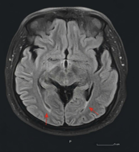 Figure 1: Focal area of loss of enhancement in the roof of one of the cystic components at the floor of the sella. A small amount of fluid showing diffusion restriction in the dependent aspect of the occipital horns, suggesting pus.
A lumbar drain was inserted to reduce CSF pressure. Subsequent CSF cultures were sterile, including at the end of intrathecal therapy.
A few days after starting antibiotic treatment, the patient’s neurological status improved, and he became responsive and able to communicate. There was defervescence of fever. The lumbar drain was removed seven days after placement, paralleled with the resolution of high opening pressure. A follow-up brain Magnetic resonance imaging after completing 21 days of intravenous antibiotics showed notable resolution of fluid/pus in the occipital horns [Figure 2]. Guided by the CSF profile [Table 1] and radiological data, intravenous meropenem was discontinued at three weeks. He was at his baseline neurologically upon discharge after a 19-day hospital stay.
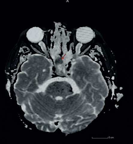 Figure 2: Follow-up brain MRI after completing 21 days of intravenous antibiotics, showing resolution of fluid/pus in the occipital horns.
Table 1: CSF analysis and gram stain.
|
Appearance
|
Cloudy
|
Clear
|
|
Leukocytes
|
5420
|
200
|
|
Polymorphs, %
|
60
|
25
|
|
Lymphocytes, %
|
40
|
75
|
|
Erythrocytes
|
47
|
0
|
|
Gram stain
|
Gram-negative rod
|
Nil
|
Intrathecal meropenem (Archifar Medochemie LTD, Cyprus) was administered at a dose of 20 mg every 12 hours, prepared in a sterile environment using aseptic technique.2 One gram of meropenem was dissolved in 20 mL of sterile water, resulting in a concentration of 50 mg/mL. Then, 0.4 mL (20 mg) of this solution was further diluted in 0.6 mL of sterile saline to make a total of 1 mL for intrathecal administration. Similarly, colistin (Atlan Pharmaceuticals, SA, Spain) was prepared for intrathecal administration at a dose of 125 000 international units once daily.3,4 One million units of colistin were dissolved in 10 mL of sterile water, 1.25 mL (125 000 IU) was taken for intrathecal administration. For intrathecal administration of antibiotics, the selected agent should always be free of preservatives and chelating agent,5 and due to the unavailability of preservatives/chelating free aminoglycosides in our institute, their use was deferred and was replaced with colistin and meropenem, both of which were ensured to be preservatives/chelating free. Written consent was obtained from the patient.
Discussion
C. koseri is a gram-negative, rod-shaped, facultative anaerobic bacterium that belongs to the Enterobacteriaceae family.1 It is commonly found in water, soil, and both human and animal digestive tracts. Despite its generally low virulence, C. koseri can cause various infections, including those of the intra-abdominal tract, urinary tract, respiratory system, skin and soft tissues, bones, bloodstream, eyes, and CNS.6
Understanding the modes of transmission is crucial in preventing the spread of C. koseri meningitis and implementing effective infection control measures. In hospital settings, transmission can occur through the use of contaminated medical equipment. Therefore, preventive measures such as proper hand hygiene and sterile techniques during invasive procedures are crucial.
Cases of C. koseri meningitis or ventriculitis in adult patients are rare, with reported instances typically involving immunocompromised individuals. Unlike our case, all reported cases (Table 27–9) were associated with some degree of immunosuppression, such as diabetes and steroid use, suggesting a potential association between C. koseri and immunosuppression, particularly in CNS involvement. Our patient, who was not diabetic or taking immunosuppressive medications, raises the likelihood of C. koseri causing meningitis in immunocompetent patients under the right circumstances.
Table 2: Reported cases of Citrobacter koseri meningitis or ventriculitis in an adult patient.
|
Booth et al,7
|
1992
|
Female, 66
|
Type II diabetes mellitus
|
*C. koseri urinary infection associated with a staghorn calculus one month before presentation.
*Spontaneous intracranial hemorrhage treated with an anterior communicating artery aneurysm clipping nine years before presentation.
|
Ampicillin
|
Gentamicin, netilmicin, cotrimoxazole, cefotaxime, and ciprofloxacin
|
Netilmicin and cefotaxime
|
ventricular drain
|
Survived/weakness and spasticity
|
|
Lind et al,8
|
2005
|
Female, 78
|
Ischemic heart disease, paroxysmal atrial fibrillation, and hypertension
|
*Ten-day course of high-dose steroid treatment
*Meningioma
|
Not available
|
Meropenem
|
Craniotomy
|
Survived/mild cognitive deficits
|
Similar to our case, one instance involved CNS surgical intervention years before presentation, suggesting a potential association between prior surgical interventions and C. koseri meningitis. This could be explained by changes in normal anatomy facilitating the entry of the organism into the brain, or the use of contaminated medical equipment during prior surgery introducing pathogenic organisms. This can increase the tendency to be infected with the same organism years later. A preceding urinary tract infection caused by the same bacteria ahead of meningitis may further suggest the possibility of hematogenous seeding.
Although C. koseri is usually sensitive to most antibiotics, it can acquire antimicrobial resistance through plasmid-mediated genes. Resistance to carbapenems in C. koseri has been reported at rates from 0 to 6.5%.1 Our case showed extended-spectrum beta-lactamase production, emphasizing the importance of individual susceptibility testing.
Despite effective antibiotic therapy, bacterial meningitis remains a serious condition with a high fatality rate. Refractory intracranial hypertension with brain herniation and brainstem compression is the most common cause of mortality in these patients.10 The use of lumbar drainage in severe acute bacterial meningitis was demonstrated to be safe and feasible. In addition to antimicrobial therapy, the use of lumbar drainage upon admission to antimicrobial therapy was significantly associated with low morbidity and mortality.11
Meningitis caused by C. koseri in neonates is associated with high rates of mortality (30%) and morbidity (50%).12 Despite all reported cases surviving, survivors may suffer from long-term neurological sequelae, such as cognitive impairment, memory problems, and motor deficits, which was observed in one case.7,12 For the same reason, it is important for healthcare providers to be aware of these potential complications and to provide appropriate follow-up care for patients who have recovered from the acute phase of the infection.
Ventriculitis is the inflammation of the ventricular fluid and the ependymal lining of the ventricles. Systemic antimicrobials based on the CSF culture results are the treatment for ventriculitis. However, the blood-brain barrier and blood-CSF barrier function as lipid layers in the CSF compartment, which may be a challenge in achieving therapeutic antimicrobial concentrations in the CNS via intravenous treatment alone. Intrathecal antibiotic treatment enables us to obtain the CSF concentration to the desired levels with a low potential for systemic toxicity. It allows direct access to the extracellular central nervous compartments by bypassing anatomical barriers, and high CSF drug levels can be attained with relatively small doses.13 Therefore; intraventricular antibiotic instillation can be used as an adjunctive therapy along with systemic antibiotics in patients with severe ventriculitis, especially with difficult to treat organisms.14
Conclusion
Meningitis caused by C. koseri is uncommon but serious in adults. The presence of previous CNS surgical intervention or immunocompromised status in a patient who presents with meningitis should raise suspicion for C. koseri as a possible cause. While C. koseri is usually susceptible to antibiotics, meropenem can be considered, especially if there is no improvement within the first 24 hours of treatment. The use of intrathecal antibiotics and lumber drainage as adjunctive therapy may contribute to patient survival.
Disclosure
The authors declare no conflicts of interest.
references
- 1. Doran TI. The role of Citrobacter in clinical disease of children: review. Clin Infect Dis 1999 Feb;28(2):384-394.
- 2. Zhang Q, Chen H, Zhu C, Chen F, Sun S, Liang N, et al. Efficacy and safety of intrathecal meropenem and vancomycin in the treatment of postoperative intracranial infection in patients with severe traumatic brain injury. Exp Ther Med 2019 Jun;17(6):4605-4609.
- 3. Metan G, Alp E, Aygen B, Sumerkan B. Acinetobacter baumannii meningitis in post-neurosurgical patients: clinical outcome and impact of carbapenem resistance. J Antimicrob Chemother 2007 Jul;60(1):197-199.
- 4. Karaiskos I, Galani L, Baziaka F, Katsouda E, Ioannidis I, Andreou A, et al. Successful treatment of extensively drug-resistant Acinetobacter baumannii ventriculitis and meningitis with intraventricular colistin after application of a loading dose: a case series. Int J Antimicrob Agents 2013 May;41(5):480-483.
- 5. LeBras M, Chow I, Mabasa VH, Ensom MH. Systematic review of efficacy, pharmacokinetics, and administration of intraventricular aminoglycosides in adults. Neurocrit Care 2016 Dec;25(3):492-507.
- 6. Indurkar SK, Mohan K, Omar BJ, Chacham S, Kumar M. Urinary tract infection in children with Nephrotic syndrome: one of the hurdles for primary care physician and pediatrician towards timely diagnosis and optimum management of the disease. J Family Med Prim Care 2022 Nov;11(11):6801-6806.
- 7. Booth LV, Palmer JD, Pateman J, Tuck AC. Citrobacter diversus ventriculitis and brain abscesses in an adult. J Infect 1993 Mar;26(2):207-209.
- 8. Lind CR, Muthiah K, Bok AP. Peritumoral Citrobacter koseri abscess associated with parasagittal meningioma. Neurosurgery 2005 Oct;57(4):E814.
- 9. Liu HW, Chang CJ, Hsieh CT. Brain abscess caused by Citrobacter koseri infection in an adult. Neurosciences (Riyadh) 2015 Apr;20(2):170-172.
- 10. Lindvall P, Ahlm C, Ericsson M, Gothefors L, Naredi S, Koskinen LO. Reducing intracranial pressure may increase survival among patients with bacterial meningitis. Clin Infect Dis 2004 Feb;38(3):384-390.
- 11. Abulhasan YB, Al-Jehani H, Valiquette MA, McManus A, Dolan-Cake M, Ayoub O, et al. Lumbar drainage for the treatment of severe bacterial meningitis. Neurocrit Care 2013 Oct;19(2):199-205.
- 12. Rodrigues J, Rocha D, Santos F, João A. Neonatal Citrobacter koseri meningitis: report of four cases. Case Rep Pediatr 2014;2014:195204.
- 13. Lee JW, Yoon Y, Kim SD, Kim YK. Intraventricular antimicrobial therapy for intractable ventriculitis: two case reports. Pediatr Infect Vaccine 2022 Apr;29(1):46-53.
- 14. Karvouniaris M, Brotis AG, Tsiamalou P, Fountas KN. The role of intraventricular antibiotics in the treatment of nosocomial ventriculitis/meningitis from gram-negative pathogens: a systematic review and meta-analysis. World Neurosurg 2018 Dec;120:e637-e650.