Cyanotic Changes of the Toes
Dawood Al-Riyami,1 Rashid Al-Sukaiti,2 Humoud Al- Duhli2
Al-Riyami D, et al. OMJ. 24, 231-233 (2009); doi:10.5001/omj.2009.47
From the 1Department of Medicine, Sultan Qaboos University Hospital 2Department of Radiology and Molecular Imaging,
Sultan Qaboos University Hospital, Muscat, Oman.
Received:
Accepted:
Address correspondence and reprint request to: Dr. Dawood Al-Riyami.
E-mail: dawood@squ.edu.om
Clinical Presentation
A 66 year old male who is known to have metastatic prostate cancer was admitted
with R-big toe pain and swelling. He reported having blue discoloration of the
toes prior to his pain. Incision and drainage of the rigA 66 year old male who
is known to have metastatic prostate cancer was admitted with R-big toe pain and
swelling. He reported having blue discoloration of the toes prior to his pain.
Incision and drainage of the right toe abscess was performed. Culture of the pus
grew Pseudomonas. On physical examination, the patient was found to have
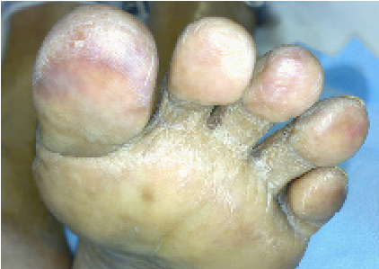
cyanotic macules of the toes in both feet (Fig.1 & 2).
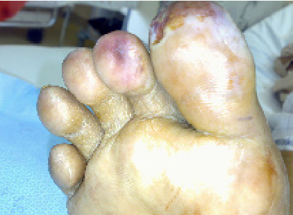
Figure 1: Right foot showing the cyanotic macules of his first and second toes pt;text-align:justify;line-height:14.0pt;text-autospace:none;vertical-align:middle;">
Figure 2: Left foot showing the cyanotic skin lesion in all of his toes The lab reports revealed significant eosinophilia. On reviewing his previous CT scan of abdomen there were circumferential soft tissue plaques involving mainly the infrarenal abdominal aorta. (Fig. 3)
Figure 3: Axial computed tomogram of the abdomen that shows circumferential soft tissue plaques with calcification involving infrarenal abdominal aorta. QUESTIONS 1. What is the differential diagnosis of the skin lesions of the toes? 2. What is the diagnosis?