
Cyanotic Changes of the Toes
Dawood Al-Riyami,1 Rashid Al-Sukaiti,2 Humoud Al- Duhli2
Al-Riyami D, et al. OMJ. 24, 231-233 (2009); doi:10.5001/omj.2009.47
From the 1Department of Medicine, Sultan Qaboos University Hospital 2Department of Radiology and Molecular Imaging,
Sultan Qaboos University Hospital, Muscat, Oman.
Received:
Accepted:
Address correspondence and reprint request to: Dr. Dawood Al-Riyami.
E-mail: dawood@squ.edu.om
Clinical Presentation
A 66 year old male who is known to have metastatic prostate cancer was admitted with R-big toe pain and swelling. He reported having blue discoloration of the toes prior to his pain. Incision and drainage of the right toe abscess was performed. Culture of the pus grew Pseudomonas. On physical examination, the patient was found to have cyanotic macules of the toes in both feet (Fig.1 & 2).
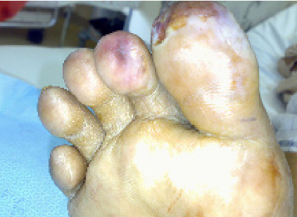
Figure 1: Right foot showing the cyanotic macules of his first and second toes
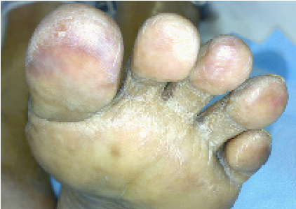
Figure 2: Left foot showing the cyanotic skin lesion in all of his toes
The lab reports revealed significant eosinophilia. On reviewing his previous CT scan of abdomen there were circumferential soft tissue plaques involving mainly the infrarenal abdominal aorta. (Fig. 3)
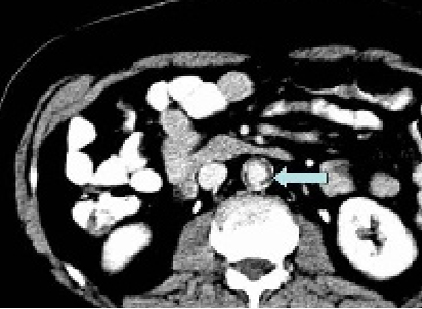
Figure 3: Axial computed tomogram of the abdomen that shows circumferential soft tissue plaques with calcification involving infrarenal abdominal aorta.
QUESTIONS
1. What is the differential diagnosis of the skin lesions of the toes?
2. What is the diagnosis?
See answers overleaf
ANSWERS
1. Antiphospholipid antibody syndrome, Vasculitis, Cholesterol emboli and Calciphylaxis.
2. Cholesterol emboli
DISCUSSION
The patient has peripheral stigmata of cholesterol emboli; the skin lesions are typical in the context of severe atherosclerosis of the aorta. Atheroemboli occur in the setting of atherosclerotic peripheral artery disease (PAD), with embolization typically developing after a procedure such as vascular or cardiothoracic surgery or arteriography. However, spontaneous embolization can occur. The ‘blue toe syndrome’ is a well recognized manifestation of atheroembolic disease.
The most common skin findings included livedo reticularis and cyanotic changes of the toes.1,2 Coexistent cutaneous signs may include, subcutaneous nodules, petechiae, purpura and hemorrhagic bullae. Atypical ischemic ulcerations of the lower extremity can arise from cholesterol emboli.3 Unlike PAD ulcerations, atypical ischemic ulcerations can evolve in the absence of significant large artery occlusive disease and thus the distal pulses are often palpable. A differential diagnosis of these unusual ischemic skin manifestation include Antiphospholipid antibody syndrome, Vasculitis and Calciphylaxis.
Multisystem organ dysfunction may be present, which often includes renal failure and hypertension. Laboratory abnormalities may include azotemia, elevated sedimentation rate, leukocytosis, eosinophilia, hypocomplementemia, thrombocytopenia, and anemia. Eosinophiluria, proteinuria, and leukocyturia can also occur. While rarely necessary, a skin or kidney biopsy will characteristically display biconvex, needle-shaped cholesterol crystal clefts within the arteriolar lumen.
ACKNOWLEDGEMENTS
The authors reported no conflict of interest and no funding has been received on this work.
REFERENCES
-
Jucgla A, Moreso F, Muniesa C, Moreno A,V idaller A. Cholesterol embolism: still an unrecognized entity with a high mortality rate, J Am Acad Dermatol. 2006; 55: 786–793
-
Kalter D.C, Rudolph A, McGavran M, Livedo reticularis due to multiple cholesterol emboli, J Am Acad Dermatol.1985; 13: 235–242.
-
Dean SM. Atypical ischemic lower extremity ulcerations: a differential diagnosis. Vasc Med. 2008 Feb;13:47-54.