
Rupture of Pseudoaneurysm of Popliteal Vessels due to Pin Migration of External Fixator
doi:10.5001/omj.2010.74
ABSTRACT
External fixators for fracture stabilization or limb lengthening cause complications such as pseudoaneurysm, acute ischemia, bleeding, compartment syndrome, and arterio venous fistula. This is a report of a patient who sustained open fracture of both bones of the right leg and the recovery period was complicated by a rare complication where migration of the proximal pin of external fixator into the popliteal vessels and rupture of pseudoaneurysm with popliteal vessels injury after a trivial trauma.
From the 1Department of Surgery, 2 Department of Orthopedics, University College of Medical Sciences & Guru Teg Bahadur Hospital, New Delhi, India.
Received: 09 May 2010
Accepted: 16 Jun 2010
Address correspondence and reprint request to: Dr. Amit Gupta, Department of Surgery, 56-A, Double Storey, Patel Nagar-II, Ghaziabad,(U.P.) India.
E-mail: dramit2411@yahoo.co.in
INTRODUCTION
External fixators provide a mechanically stable environment to allow fracture stabilization or limb lengthening. Most of the complications are related to pin or wire insertion. The complications include pseudoaneurysm, acute ischemia, bleeding, compartment syndrome, and arteriovenous fistula. Here we report a rare and very interesting case of a 35 years old male who sustained open fracture of both bones of his right leg at the junction of upper one third and middle third level with consequent infection and bone loss. The recovery period was complicated by a rare complication of migration of proximal pin of external fixator into the popliteal vessels and rupture of pseudoaneurysm with popliteal vessels injury after a trivial trauma.
CASE REPORT
A 35 years old male presented in the orthopedics emergency with complaints of pain and swelling in the right knee and leg for the last two days after a trivial trauma with external fixator in situ in the right leg which was positioned three months back for fracture of both bones in his right leg at the junction of upper one third and middle third level following road traffic accident, but patient did not turn up for follow up after being discharged from the previous admission.
On general physical examination, vitals were stable and there was no pallor. Local examination of lower right limb revealed swelling in popliteal region and medial aspect of upper one third of his right leg with external fixator in situ. The right lower limb was cold to touch below the level of swelling. The popliteal artery, posterior tibial and dorsalis pedis artery pulsations were not palpable. There was no neural deficit. The left lower limb was unremarkable on examination. An X-ray of the right knee and leg showed posterior migration of proximal pin of external fixator. (Fig. 1)
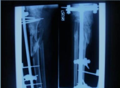
Figure 1: X-ray showing posterior migration of proximal pin of external fixator.
Color Doppler right lower limb showed absent blood flow at the level of popliteal artery and below. The diagnosis of compartment syndrome with popliteal vessel injury and possibility of pseudoaneurysm rupture with external fixator in situ was made. The patient was scheduled for emergency exploration under general anaesthesia. The external fixator was removed and liberal fasciotomy was performed. On exploration of the right popliteal fossa and leg, there was a large clot causing clot tamponade in the popliteal fossa with ruptured pseudoaneurysm in the lateral wall along with longitudinal tear of 2 cm in the anterior wall of the popliteal artery 5 cm below pseudoaneurysm with shattered popliteal vein. (Figs. 2a & 2b)
The clot was removed and the popliteal vein was ligated. The ruptured pseudoaneurysm was excised and the defect in arterial wall was repaired with prolene 5-0. Thrombectomy by 6 Fr. Fogarty’s catheter and repair of popliteal artery tear with prolene 5-0 was done. POP cast was applied for immobilization of the fracture segment.
On the second postoperative day, the limb was warm and pulsation in the right dorsalis pedis artery was palpable. Postoperative recovery was along expected lines except wound infection on fifth postoperative day for which serial debridement was done under local anaesthesia and the wound was managed by saline dressings and antibiotics.
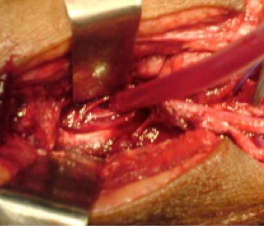
Figure 2A: Longitudinal tear in popliteal artery.

Figure 2B: Shattered popliteal vein and ruptured pseudoanuerysm.
DISCUSSION
Pseudoaneurysm of the popliteal artery may be secondary to several factors, trauma being the predominant cause. Pseudoaneurysm of the popliteal artery represents approximately 1% of all vascular lesions, with a high rate of limb loss due to thromboembolic complications.1,2 Several mechanisms of trauma are involved. Penetrating traumas due to stab wounds or gunshot wounds, and femoral or tibial fractures or fracture/luxation may cause direct lesion to the vessel.
Blunt traumas without fractures rarely lead to pseudoaneurysms, given that only a great mechanical force can cause trauma to the popliteal artery. The popliteal fossa is surrounded by a highly resistant structure of muscles and bones; thus, mild traumas rarely lead to arterial damage without damaging surrounding structures.
Structural alterations on the arterial wall, such as those caused by collagen or connective tissue diseases and by arteritis secondary to septic embolism in patients with infectious endocarditis can rarely cause popliteal pseudoaneurysm formation. Such diseases invariably lead to structural damage, weaken the arterial wall and make the artery more vulnerable to spontaneous ruptures or ruptures due to mild traumas.
The causative agent in this patient was the damage caused by migration of the pin. External fixators provide a mechanically stable environment to allow fracture stabilization or limb lengthening. Most complications are related to pin or wire insertion. The incidence of neurovascular injury associated with placement of external fixator half-pins has been quoted as 0.6% as reported by Green in a review of over 4,000 injuries treated by external fixation.3
This incidence has remained low because of adherence to principles of external fixation for avoidance of neurovascular injury as described by Behrens and Searls in 1986. Other complications include false aneurysm, acute ischemia, bleeding, compartment syndrome, and arteriovenous fistula. The first theory to explain the etiology of false aneurysms in relation to external fixation was direct vascular damage from the inappropriate position of pins or wires resulting in clinical symptoms within days or weeks.4 It is remarkable that pseudoaneurysms are reported following pin or wire withdrawal as well.5
In order to explain this, as well as the late appearance of some lesions, the theory of vascular lesion due to erosion from contact with a pin or a wire has been introduced.6 This is supported by a study by Dwyer who found that it is almost impossible to pierce any major vessel with the pins.7 Another cause of pseudoaneurysm formation that has been reported is bone distraction. Lengthening may either aggravate an unrecognized surgical arterial injury or precipitate de novo rupture of an artery scarred from a previous trauma.8
We were able to find only 7 cases in the literature of arterial pseudoaneurysm related to application of external fixation to the tibia, in which one was affecting the distal popliteal artery. The popliteal artery is more commonly injured when fixators are placed at the femur.9 This case was unique because not only was there pseudoaneurysm rupture, but also popliteal artery tear. Clinical symptoms are presented early if there is an immediate arterial injury or late if the lesion is due to erosion from impingement.10 The symptoms include bleeding through the pin holes, lowering of hematocrit level, appearance of a mass (often pulsating), and sometimes pain.
Possible complications from a false aneurysm include bleeding, occlusion and ischemia, rupture and compartment syndrome, and infection from contamination by an infected pin.11 Once injury is identified, several approaches to the management of popliteal artery injury have been described. Reversal of ischemia (if present) is of the utmost importance. Severe ischemia has been shown to be a strong predictor of poor outcomes with regard to limb amputation and sensory deficits. Strict follow-up of orthopaedic trauma is therefore essential.
CONCLUSION
A high index of suspicion and awareness can lead to early diagnosis and treatment of pseudoaneurysms of the popliteal artery and prevent the serious complications associated with these lesions. Although our patient presented after 48 hours of popliteal vessel injury, we were able to salvage the limb because of timely intervention and aggressive management.
ACKNOWLEDGEMENTS
The authors reported no conflict of interest and no funding was received on this work.
References
-
Marcove RC, Lindeque BG, Silane MF. Pseudoaneurysm of the popliteal artery with an unusual arteriographic presentation. A case report. Clin Orthop 1988; 234:142-4.
-
Gillespie DD, Cantelmo NL. Traumatic popliteal artery pseudoaneurysm: case report and review of the literature. J Trauma 1991;31(3):412-15.
-
Topp R, Hayda R, Benedetti G, Twitero T, Carmack DB. The Incidence of Neurovascular Injury during External Fixator Placement without Radiographic Assistance for Lower Extremity Diaphyseal Fractures: A Cadaveric Study.The Journal of Trauma: Injury, Infection, and Critical Care: 2003;55 (5): 955-958.
-
Rich NM, Hobson RW II, Collins GJ Jr. Traumatic arteriovenous fistulas and false aneurysms: a review of 558 lesions. Surgery. 1975; 78(6):817-828.
-
Polak WG, Pawlowski S, Skora J, Morasiewicz L, Janczak D, Oleszkiewicz M, et al. Vascular complications after the treatment with Ilizarov external fixators. VASA. 2001; 30(2):138-140.
-
Waldhausen J, Mosca V, Johansen K, Schaller R. Delayed presentation of popliteal artery injury during Ilizarov limb lengthening. Orthopedics. 1998; 21(4):477-478.
-
Dwyer NS. Preliminary report upon a new fixation device for fractures of long bones. Injury. 1973; 5(2):141-144.
-
Griffith JF, Cheng JC, Lung TK, Chan M. Pseudoaneurysm after high tibial osteotomy and limb lengthening. Clin Orthop Relat Res. 1998; (354):175-179.
-
Sharp RJ, Latham JM, Simpson AH. Post operative swelling in Ilizarov leg lengthening. Injury. 1998; 29(9):717-718.
-
Braito W, Montanari C, Caracciolo F, Paroni G, Domenella G. False aneurysm of the anterior tibial artery in lower leg fractures treated with the Ilizarov external fixator. Case report. Italian Journal of Orthopaedics and Traumatology. 1992; 18(1): 135-139.
-
Paul MA, Patka P, van Heuzen EP, Koomen AR, Rauwerda J. Vascular injury from external fixation. Case reports. J Trauma. 1992; 33(6):917-920.
How to cite this URL
Gupta A, Tandon A, Quereshi AQ, Kumar S, Arora S. Rupture of Pseudoaneurysm of Popliteal Vessels due to Pin Migration of External Fixator. OMJ [Online] 2010 July; 25(3). Available at http://www.omjournal.org/CaseReports/FullText/201007/FT_9RuptureofPseudoaneurysmofPopliteal.html.