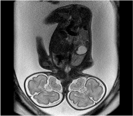
The antenatal magnetic resonance imaging (MRI) of a third-trimester primipara patient showed a fetus in a vertical position with two separate heads, a common thorax, abdomen, and pelvis with a single pair of upper and lower limbs [Figure 1]. Both heads appeared equal and symmetric with normal brain development [Figure 2]. Two separate upper cervical spinal cords fused to form a single spinal cord in the mid/lower cervical spine level. There was a single heart present in the thoracic cavity. The intra-abdominal organs appeared normal in position, structure, and number [Figure 1]. The findings were confirmed at delivery [Figure 3].
Questions
- What is this variety of conjoint twins called?
- Is there any associated cardiac anomaly present in this case?
- What is the investigation of choice for evaluation of cardiac anomalies?
Answers
- Dicephalic parapagus twins.
- Dextrocardia.
- Fetal echocardiography.
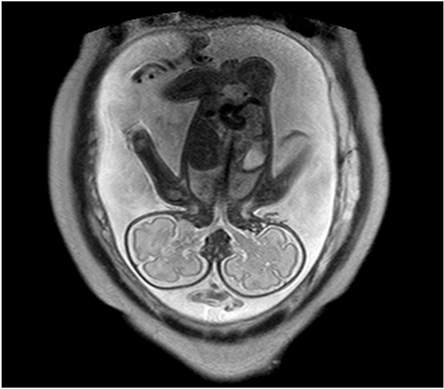
Figure 1: Coronal T2-weighted turbo spin echo (TSE) showed the fetus in a vertical position with two separate heads, two cervical spinal cords fusing at the mid-cervical spine level, a common thorax, abdomen, and pelvis, and a single pair of upper and lower limbs and a single umbilical cord.
![]()
Figure 2: Coronal T2-weighted turbo spin echo (TSE) the posterior half of the fetal heads showed normally and bilaterally symmetric development of the fetal brains on both sides. Normal appearing cerebral and cerebellar hemispheres, with normal sulci and gyri, were noted.
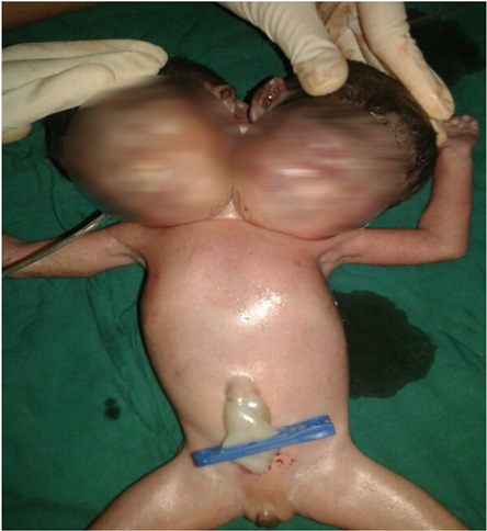
Figure 1: Coronal T2-weighted turbo spin echo (TSE) showed the fetus in a vertical position with two separate heads, two cervical spinal cords fusing at the mid-cervical spine level, a common thorax, abdomen, and pelvis, and a single pair of upper and lower limbs and a single umbilical cord.
Discussion
A dicephalic parapagus twin is an extremely rare entity with very limited literature describing the condition. The incidence of conjoined twins is one in 250,000 live births with 3:1 female preponderance.1 Conjoined twinning is a random event not related to maternal age, parity, race, and heredity and is explained variedly by fission and fusion theories.2 The widely accepted fission theory proposes that conjoined twins occur when a fertilized ovum begins to split into identical twins but is interrupted during the process and develops into two partially formed individuals who are stuck together.3 The fusion theory postulates that two monovular embryonic discs may lie adjacent to one another at various angles, and may become secondarily united homologously.4
The present case was diagnosed at an advanced gestational age due to the late primary presentation, but similar cases can be diagnosed as early as the tenth week of gestation.5 Ultrasound and fetal MRI form the mainstay of diagnosis in such cases. Ultrasound is more useful at the beginning of pregnancy and for initial detection, with the ability to detect conjoint twinning around 12 weeks. However, it has limited use in defining the extent, exact site and anatomical details of fusion, which determine the prognosis and further management.
Ultrafast short duration T2-weighted MRI sequences provide excellent image quality, enabling fetal MRI to provide excellent anatomical details, extent, level of fusion and spatial relationship even in the presence of severe oligohydramnios, maternal obesity, and advanced gestational age. Therefore scoring significantly over ultrasound.1 Ultrafast imaging sequence MRI is helpful to corroborate and refine ultrasound diagnoses. Prenatal MRI facilitates perinatal management, and family counseling should be advised in cases diagnosed with conjoint twins on ultrasound. Fetal MRI is also a valuable adjunct to ultrasound before fetal surgical interventions for selected life-threatening birth defects or planning emergent separation at birth, either of which can be life saving.6,7
Prenatal diagnosis can help guide decisions so that both fetal and maternal morbidity and mortality can be minimized. Classification of conjoined twins is paramount for guiding obstetrical management. A vast majority of conjoined twins do not survive the first 24 hours of life. Therefore, the fetal chance for survival has to be weighed against the potential surgical morbidity to the mother and the feasibility of vaginal delivery.8
The postnatal prognosis of conjoint twins depends on the extent of fusion.9 In lateral fusion (i.e. parapagus) there is a common abdomen, pelvis and lower extremity with a variable number of upper limbs. While the term dicephalic parapagus implies two entirely separate heads, faces, and a single heart, two hearts have also been reported. In the presence of a single heart, surgical separation is rarely successful. In those with two hearts, survival is largely dependent on abnormalities of other viscera.10 The site and the extent of the union are reflected in the varying degrees of involvement of the viscera, particularly the heart, diaphragm, gastrointestinal tract, and the urogenital system. Generally, dicephalic twins have two oesophagi, which may join in the middle of the thorax, or both may empty into a single stomach.11
When serious malformations are diagnosed in cases of dicephalic twins, which are incompatible with postnatal life at early stages of gestation, the parents should be counseled regarding the outcome and early termination of such pregnancies. Imaging by either ultrasound or MRI is helpful in deciding whether termination of pregnancy is advisable.12
Conclusion
Prenatal diagnosis using ultrasound and antenatal MRI plays a vital role in classification, prognostication, and management of complex conjoint twinning. An integrated approach including a radiologist, obstetrician, pediatric surgeon, and neonatologist should be practiced in deciding the viability, mode of delivery, and postnatal care required.
Disclosure
The authors reported no conflicts of interest. The parents of the infant gave their consent for the publication of the image in this case report.
Acknowledgements
I am deeply thankful to Dr. Bhupendra Bhandari for postnatal images, and to Dr. Jalpa Daftary for her guidance.
references
- McHugh K, Kiely EM, Spitz L. Imaging of conjoined twins. Pediatr Radiol 2006 Sep;36(9):899-910, quiz 1002-1003.
- Kaufman MH. The embryology of conjoined twins. Childs Nerv Syst 2004 Aug;20(8-9):508-525.
- Bondeson J. Dicephalus conjoined twins: a historical review with emphasis on viability. J Pediatr Surg 2001 Sep;36(9):1435-1444.
- Spencer R. Theoretical and analytical embryology of conjoined twins: part I: embryogenesis. Clin Anat 2000;13(1):36-53.
- Vural F, Vural B. First trimester diagnosis of dicephalic parapagus conjoined twins via transvaginal ultrasonography. J Clin Ultrasound 2005 Sep;33(7):364-366.
- Quinn TM, Hubbard AM, Adzick NS. Prenatal magnetic resonance imaging enhances fetal diagnosis. J Pediatr Surg 1998 Apr;33(4):553-558.
- Mackenzie TC, Crombleholme TM, Johnson MP, Schnaufer L, Flake AW, Hedrick HL, et al. The natural history of prenatally diagnosed conjoined twins. J Pediatr Surg 2002 Mar;37(3):303-309.
- Vaughn TC, Powell LC. The obstetrical management of conjoined twins. Obstet Gynecol 1979 Mar;53(3)(Suppl):67S-72S.
- Başaran S, Güzel R, Keskin E, Sarpel T. Parapagus (dicephalus, tetrabrachius, dipus) conjoined twins and their rehabilitation. Turk J Pediatr 2013 Jan-Feb;55(1):99-103.
- McMahon CJ, Spencer R. Congenital heart defects in conjoined twins: outcome after surgical separation of thoracopagus. Pediatr Cardiol 2006 Jan-Feb;27(1):1-12.
- Spencer R. Theoretical and analytical embryology of conjoined twins: part II: adjustments to union. Clin Anat 2000;13(2):97-120.
- Owolobi AT, Oseni SB, Sowande OA, Adejuyigbe O, Komolafe EO, Adetiloye VA, et al. Dicephalus dibrachius dipus conjoined twins in a triplet pregnancy. Trop J Obstet Gynaecol 2005;22:87-88.