|
Abstract
The presence of echogenic amniotic fluid on sonography is uncommon and its clinical significance is not well appreciated. Very echogenic amniotic fluid has been attributed to meconium, blood, or vernix caseosa. Two cases of patients with echogenic amniotic fluid at term are presented here. In the first case, the patient’s management was altered as the finding of echogenic amniotic fluid was interpreted to be thick meconium. The second case was induced for post-datism and the amniotic fluid was found clear during labour. Since the first reported cases of meconium with echogenic amniotic fluid on sonography by Benacerraf et al. (1984), larger studies have consistently shown that echogenicity is not predictive of meconium. As with our cases, meconium was suspected in both patients with dense echogenic amniotic fluid. Labor was induced in the first case to avoid fetal distress and asphyxia with increasing gestation. Meconium was not present in either of our cases and both the babies were healthy.
Keywords: Prenatal ultrasound; Echogenic amniotic fluid; Thick meconium; vernix; Term-pregnancy.
Introduction
The presence of echogenic amniotic fluid on sonography is uncommon and its clinical significance is not well appreciated. Very echogenic fluid has been attributed to meconium, blood or vernix caseosa. Finding meconium in the amniotic fluid raises concerns about fetal well-being, the ability of the fetus to tolerate labor, or the risk of intrauterine fetal demise.1 Therefore, when interpreting the sonographic findings of echogenic amniotic fluid, it is important to determine whether or not it represents meconium, as management may be affected with its detection. Early literature supported the identification of meconium with the finding of echogenic amniotic on sonography.2 However, more recent studies and case reports have reported findings of vernix rather than meconium by amniocentesis or at delivery. This is a case report of two patients who presented with echogenic amniotic fluid at term. In the first case, the patient’s management was altered as the finding of echogenic amniotic fluid was interpreted to be thick meconium. The findings of our cases agree with the more recent literature that sonographically echogenic amniotic fluid at term gestation is most likely due to vernix.
Case 1
A 35-year-old primigravida was followed at the high risk clinic for a history of two previous cone biopsies for cervical dysplasia. A Macdonald cerclage was inserted at 13 weeks, and then removed at 36 weeks for cervical incompetence. Pregnancy was otherwise uneventful. At 39 weeks of gestation, obstetric ultrasound findings showed extremely dense echogenic amniotic fluid (Figs. 1 and 2). The amniotic fluid index measured 17.9 cm, adequate fetal growth, normal umbilical artery Doppler, and a normal biophysical profile. There was concern that the echogenicity of the amniotic fluid represented thick meconium. Although fetal monitoring was reassuring, the findings of calcified placenta and possible meconium-stained amniotic fluid led to the decision to induce labor. Artificial rupture of membranes was performed and the amniotic fluid was clear. Prolonged second stage of labor and non-reassuring heart tracing resulted in emergency cesarian section. At cesarian section, the amniotic fluid was clear, baby’s Apgar scores were 8 at 1 minute and 9 at 5 minutes. Baby weighed 3510 g with cord pH of 7.3 and base excess of -5.2. Mother and baby were discharged in good condition three days post-operatively.
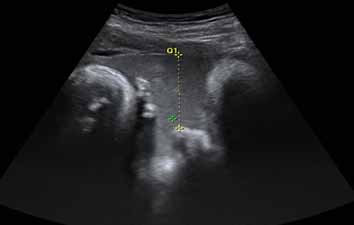
Figure 1: Obstetric ultrasound of patient 1 showing very dense echogenic amniotic fluid.
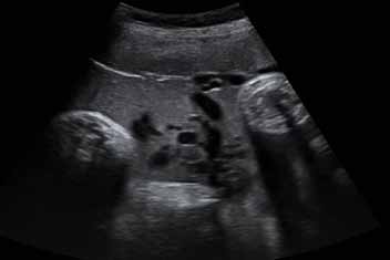
Figure 2: Obstetric ultrasound of pateint 1 showing very dense echogenic amniotic fluid with umbilical cord loops.
Case 2
A 29-year-old gravida 2, para 1, presented at 41 weeks and 3 days for induction of labor for post-dates. An ultrasound done at 41 weeks reported very dense amniotic fluid (Fig. 3). She was induced with IV oxytocin. Membranes were ruptured spontaneously in active labor and the amniotic fluid was clear. She delivered a healthy baby boy, weighing 3812 g, with Apgar score of 9 at 1 minute and 9 at 5 minutes. Cord pH was 7.4 and base excess was 1.6. Mother and baby were discharged in good condition two days post-delivery.
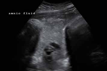
Figure 3: Obstetric ultrasound of patient 2 showing very dense echogenic amniotic fluid.
Discussion
Vernix caseosa is a complex fatty substance derived from the desquamated epithelial cells and sebaceous material.3 In 1978, Bree identified vernix in the amniotic fluid during the late third trimester by the presence of small solid particles on sonography.4 This was confirmed by amniocentesis and examination of the fluid at delivery. This finding was in agreement with an article by Verpoest et al.5 who developed a macroscore to explain the changes in appearance of amniotic fluid during pregnancy. They noted that the flakes of vernix appear only after 36 weeks and cause clouding of the fluid, which increases toward term. Khaleghian reported in 1983 the second case of echogenic particles within the amniotic fluid in the second trimester.6 However, blood was found to be the most likely source of the finding.
The first identification and description of meconium-stained amniotic fluid on ultrasound was reported by Benacerraf et al. in 1984.2 Two patients underwent evaluation of fetal well-being at 42 weeks and were both found to have amniotic fluid containing low-level echoes with accentuation of the echoes in the dependent areas. The fluid in the fundus and anterior portions of the uterus was clear and almost free of echoes, while in the dependent portions of the uterus, the contrast between the anechoic appearance of the umbilical vein and the surrounding echogenic amniotic fluid was easily seen. Due to the finding of dense echoes clustering in the dependent portions of the uterus, they concluded that this was more consistent with meconium as vernix tends to produce more diffuse and less dense low-level echoes. Therefore meconium-stained amniotic fluid can be detected in antepartum by the following findings on ultrasound: diffuse echogenic pattern throughout the amniotic cavity, a clear contrast between the amniotic fluid and the umbilical cord, and layering in the more dependent areas.2
In contrast, more recent studies and case reports on echogenic amniotic fluid have reported findings of vernix rather than meconium by amniocentesis or at delivery. DeVore and Platt presented a case of sludge-like amniotic fluid on ultrasound,7 which had the appearance of what was reported by Benacerraf et al.2 to be associated with meconium. Because of the possibility of fetal distress, the patient was induced into labor. Artificial rupture of the membranes demonstrated amniotic fluid with thick vernix and no evidence of meconium and the baby was unaffected. Two other case reports with similar echogenic findings demonstrated the presence of vernix on amniocentesis and good baby outcomes.3,8
In an interesting study by Giacomello in 1988, biophysical profiles of 1100 patients were examined with only one case of a "dense" AF pattern, found in a primigravida at 38 weeks.9 The fluid was described to be so echogenic in dependent areas that the umbilical cord looked like a fossil. No invasive assessment of the fluid was done, but repeated biophysical variables were done to monitor fetal well-being. The pregnancy was allowed to term, a healthy baby was delivered, and abundant vernix was identified in the amniotic fluid at birth. Subsequently, Sepulveda et al.8 in Chile conducted a literature review of the above mentioned cases, and recommended that if echogenic amniotic fluid is identified on sonography, meconium should be ruled-out by amniocentesis or amnioscopy, or fetal well-being should be assessed by non-stress test or biophysical profile, in order to avoid false positive diagnoses of fetal distress leading to unnecessary precipitation of delivery.
Since these early case reports, larger prospective and retrospective studies have been conducted to further examine the clinical relevance of echogenic amniotic fluid antenatally. A prospective study by Sherer et al.10 reported a sensitivity of 100%, a specificity of 69%, a positive predictive value of 10%, and a negative predictive value of 100% in detecting meconium when homogenous echogenic AF is seen on sonography. A retrospective study by Brown et al.11 showed out of 19 cases, vernix was present in 18 (95%) patients and meconium in one (5%). They concluded that the ultrasound finding of echogenic amniotic fluid is not a reliable indicator of meconium or blood, and therefore should not typically alter antenatal management. More recently, Petrikovsky et al.12 studied prospectively 19 twin pregnancies with which the amniotic fluid in one sac was anechoic and that in the other sac was echogenic. The amniotic fluid was then assessed clinically by amniocentesis or upon delivery within 48 hours after sonography. They found that in the twins with echogenic fluid, 32% (6) had clear fluid, 63% (12) had vernix caseosa, and 5% (1) had meconium. In the co-twin with anechoic fluid, 47% (9) had clear fluid, 32% (6) had vernix caseosa, and 21% (4) had meconium. They concluded that echogenicity on prenatal sonography is not predictive of meconium.
In the most recent large prospective study in 2005, Mungen et al.1 studied the relationship between pregnancy outcome with echogenic amniotic fluid at term of 950 women with singleton pregnancies at >37 weeks of gestation. Echogenic amniotic fluid was found in 7% of the women. There was no statistically significant difference (p=0.79) between the incidences of meconium-stained amniotic fluid at delivery with or without echogenic amniotic fluid (11% and 10%, respectively). They also found that there were no statistically significant differences in rates of adverse pregnancy outcomes between the echogenic and anechoic, which included outcomes such as preeclampsia/eclampsia, diabetes, puerperal infection, cesarean delivery, Apgar scores <7 at 1 and 5 minutes, small for gestational age, and neonatal intensive care admission. They concluded that since sonographically echogenic amniotic fluid at term gestation is not associated with any adverse pregnancy outcome, such a finding should not alter routine antenatal management.
Since the first reported cases of meconium with echogenic amniotic fluid on sonography by Benacerraf et al.2 in1984, larger studies have consistently shown that echogenicity is not predictive of meconium. As with our cases, meconium was suspected in both patients with dense echogenic amniotic fluid. Labor was induced in the first case to avoid fetal distress and asphyxia with increasing gestation. Meconium was not present in either of our cases and both the babies were healthy.
Conclusion
Our findings are consistent with the literature that echogenic amniotic fluid on ultrasound most likely reflects the presence of vernix rather than meconium in 60% to 95% of cases. Upon reviewing the literature, the decision to induce the first case at 39 weeks with a reassuring heart rate should not have been influenced by the finding of thick echogenic amniotic fluid on sonography.
We are in agreement with the recommendation that sonographic detection of echogenic amniotic should not be immediately interpreted as meconium and that such a finding warrants immediate non-invasive monitoring (non-stress test and biophysical profile) to assess fetal well-being. If the assessment is reassuring, routine antenatal management does not have to be altered and delivery can be considered if indicated otherwise. If it is non-reassuring, management can be determined by weighing the risks and benefits of amniocentesis versus delivery.
Acknowledgements
The authors reported no conflict of interest and no funding was received for this work.
References
1. Müngen E, Tütüncü L, Muhcu M. Pregnancy outcome in women with echogenic amniotic fluid at term gestation. Int J Gynaecol Obstet 2005 Mar;88(3):314-315.
2. Benacerraf BR, Gatter MA, Ginsburgh F. Ultrasound diagnosis of meconium-stained amniotic fluid. Am J Obstet Gynecol 1984 Jul;149(5):570-572.
3. Hill LM, Breckle R. Vernix in amniotic fluid: sonographic detection. Radiology 1986 Jan;158(1):80.
4. Bree RL. Sonographic identification of fetal vernix in amniotic fluid. J Clin Ultrasound 1978 Aug;6(4):269-270.
5. Verpoest MJ, Seelen JC, Westerman CF. Changes in appearance of amniotic fluid during pregnancy - the macroscore. J Perinat Med 1976;4(1):12-25.
6. Khaleghian R. Echogenic amniotic fluid in the second trimester: a new sign of fetal distress. J Clin Ultrasound 1983 Nov-Dec;11(9):498-501.
7. DeVore GR, Platt LD. Ultrasound appearance of particulate matter in amniotic cavity: vernix or meconium? J Clin Ultrasound 1986 Mar-Apr;14(3):229-230.
8. Sepúlveda WH, Quiroz VH. Sonographic detection of echogenic amniotic fluid and its clinical significance. J Perinat Med 1989;17(5):333-335.
9. Giacomello F. Sonographic findings of dense amniotic fluid. Am J Obstet Gynecol 1988 May;158(5):1242-1243.
10. Sherer DM, Abramowicz JS, Smith SA, Woods JR Jr. Sonographically homogeneous echogenic amniotic fluid in detecting meconium-stained amniotic fluid. Obstet Gynecol 1991 Nov;78(5 Pt 1):819-822.
11. Brown DL, Polger M, Clark PK, Bromley BS, Doubilet PM. Very echogenic amniotic fluid: ultrasonography-amniocentesis correlation. J Ultrasound Med 1994 Feb;13(2):95-97.
12. Petrikovsky B, Schneider EP, Gross B. Clinical significance of echogenic amniotic fluid. J Clin Ultrasound 1998 May;26(4):191-193.
|