|
Introduction
Small intestinal necrosis followed by gangrene may occur due to intestinal obstruction or ischemic bowel disease. Bowel obstruction in pregnancy is not common compared to the general population. The conditions commonly precipitating intestinal obstruction include adhesions from previous pelvic operation including lower uterine cesarean section (LUCS), cecal and small bowel volvulus, and Meckel’s diverticulum.1,2 Ischemic bowel disease is also uncommon and may be caused by acute or chronic conditions. Acute intestinal ischemia is caused by arterial occlusion, venous thrombosis, vasculitis (including systemic lupus erythematosus [SLE], polyarteritis nodosa), embolism and non-occlusive disease. Venous thrombosis occurs in portal hypertension, inflammatory and neoplastic conditions, and hypercoagulable states (pregnancy, protein C and S deficiency). Chronic intestinal ischemia, known as abdominal angina is an uncommon condition, associated with atherosclerotic vascular disease.3 This report describes a rare case of identified idiopathic segmental small gut gangrene in an elderly primigravida with term size pregnancy who underwent lower uterine cesarean section for fetal distress.
Case Report
A 32-year-old primigravida with term pregnancy presented at the emergency department with acute pain in the abdomen for the last 12 hours. The patient was conscious and hemodynamically stable. There was guarding and tenderness over the whole abdomen and fetal distress, but there was no fever, vomiting or constipation. The patient had blood pressure of 120/80 mmHg and pulse 85/min. Examination of abdomen revealed fundal height of 32 weeks, no operative scar mark, and fetal heart sound (FHS) was 190/min. Vaginal examination revealed an os of 2 cm with the membrane intact and stationed high up. Laboratory investigations revealed Hb% of 12 gm%, blood sugar (post prandial) of 75 mg/dL. HBsAg was negative, VDRL was also negative, free T4 and TSH levels were within normal limits. Ultrasonography (USG) showedaverage liquor amnii and a viable fetus of approximately 32-week size with cephalic presentation.
LUCS was done under general anesthesia for fetal distress and unexpectedly, an approximately 15 cm long segment of ileum, 3 cm away from the ileocecal junction was found to be gangrenous (Fig. 1). Resection of the gangrenous segment was done and proximal-end ileostomy and distal mucous fistula was performed to avoid leakage in an unprepared bowel. Postoperatively, the patient was doing well and sent home after one month of delivery. The delay in discharge was due to a postoperative wound dehiscence. Postoperative laboratory evaluation did not reveal any coagulation abnormality. The patient’s prothrombin time was 13.5 seconds (control: 13 seconds); activated partial thromboplastin time was 34 seconds (control: 35 seconds), and fibrinogen level was 256 mg/dL (normal: 233-496 mg/dL). Screens for antiphospholipid antibodies like lupus anticoagulant and anti-cardiolipin antibodies were negative (anti-cardiolipin antibody: patient’s IgG value = 6 GPL [normal range = 0-15 GPL]; patient’s Ig M value = 7 MPL [normal range = 0-15 MPL]). Inherited thrombotic disorders like deficiency of protein S (patient’s value = 78%; normal range = 70% to 140%), protein C (patient’s value = 101%; normal range = 70% to 140%), antithrombin III (patient’s value = 95.5%; normal range = 70% to 130%), and factor V Leiden mutation (activated protein C resistance, ratio: 2.2 in the patient, normal level ≥2.1) which demonstrated negative results.
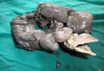
Figure 1: Gangrenous part of small gut removed during LUCS.
Discussion
In this case, gangrene was found approximately 15 cm in length at the terminal part of the ileum, 3 cm away from the ileocecal junction. Preoperatively, there were no adhesions and bands, Meckels’ diverticulum or volvulus, which are the usual causes of gangrene of the small intestine even during pregnancy.1,2 Acute intestinal ischemia is caused by arterial occlusion, venous thrombosis, vasculitis (SLE, polyarteritis nodosa), antiphospholipid syndrome, embolism and non-occlusive diseases. Venous thrombosis occurs in portal hypertension, inflammatory and neoplastic conditions and hypercoagulable states such as pregnancy, antiphospholipid syndrome, factor V Leiden mutation etc.4,5
Patients with acute ischemia have severe abdominal pain with few physical, laboratory or X-ray findings. As necrosis progresses, there is peritonitis with abdominal distention, ileus, restlessness and anxiety. Pregnancy itself is a hypercoagulable state which rarely precipitates deep vein thrombosis (DVT) and venous gangrene.6 Two recent case reports demonstrated idiopathic mesenteric vein thrombosis in pregnancy suggesting a hypercoagulable state.7,8
Conclusion
The present case is atypical due to the absence of the usual causes of small gut gangrene in pregnancy. Pregnancy itself was suggested to be the cause of this gangrene due to its hypercoagulable state;however, no hypercoagulability persisted during the patient’s follow-up after postpartum period.
Acknowledgements
The authors reported no conflict of interest and no funding was received for this work.
References
1. Essilfie P, Hussain M, Stokes IM. Small bowel infarction secondary to volvulus during pregnancy: a case report. J Reprod Med 2007 Jun;52(6):553-554.
2. Rudloff U, Jobanputra S, Smith-Levitin M, Kessler E. Meckel’s diverticulum complicating pregnancy. Case report and review of the literature. Arch Gynecol Obstet 2005 Jan;271(1):89-93.
3. Tyson RL. Diagnosis and treatment of abdominal angina. Nurse Pract 2010 Nov;35(11):16-22, quiz 22-23.
4. Robertson L, Wu O, Langhorne P, Twaddle S, Clark P, Lowe GD, et al; Thrombosis: Risk and Economic Assessment of Thrombophilia Screening (TREATS) Study. Thrombophilia in pregnancy: a systematic review. Br J Haematol 2006 Jan;132(2):171-196.
5. Ruiz-Irastorza G, Crowther M, Branch W, Khamashta MA. Antiphospholipid syndrome. Lancet 2010 Oct;376(9751):1498-1509.
6. James AH. Pregnancy and thrombotic risk. Crit Care Med 2010 Feb;38(2)(Suppl):S57-S63.
7. Lin H, Lin CC, Huang WT. Idiopathic superior mesenteric vein thrombosis resulting in small bowel ischemia in a pregnant woman. Case Reports in Obstetrics and Gynecology. vol. 2011, Article ID 687250, 3 pages, 2011. doi:10.1155/2011/687250.
8. Salati SA, Rather AA. Primary mesenteric vein thrombosis in pregnancy. Journal of symptoms and sign 2012; 1:e1-e3.
|
|