A 44-year-old Omani male presented to the Dermatology Department with a four-month history of skin lesion over the face, associated with a feeling of tightness. He reported no history of itching, oral ulceration, fever, shortness of breath, or eye symptoms. There was no history of photosensitivity. His medical and drug history were unremarkable.
General physical and systemic examinations were normal. Local examination revealed an erythematous plaque measuring about 2 × 3 cm with superficial scales and atrophic violaceous center over the left cheek [Figure 1]. A similar smaller lesion was noted over the distal dorsum of the nose.
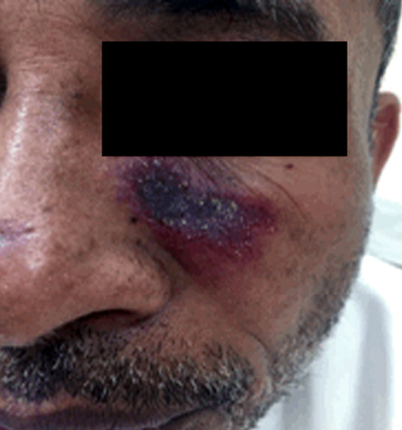
Figure 1: Clinical picture of the patient.
Complete blood count and blood biochemistry were normal. Antinuclear antibodies test was negative. His chest X-ray was normal and the Mantoux test was negative. A skin punch biopsy was done, which revealed epidermal atrophy, follicular plugging, and basal layer vacuolization. The dermis showed perivascular and perifollicular lymphocytic infiltrate. Staining for acid-fast bacilli was negative.
Question
- What is the diagnosis?
a. Actinic lichen planus.
b. Cutaneous leishmaniasis.
c. Cutaneous sarcoidosis.
d. Discoid lupus erythematosus.
e. Lupus vulgaris.
Answer
d. Discoid lupus erythematosus.
A diagnosis of discoid lupus erythematosus (DLE) was made based on clinical and histopathological features. The patient was treated with regular sunblock and potent topical steroids but showed mild response after two months, so he was started on hydroxychloroquine.
Discussion
DLE is the most common form of chronic cutaneous lupus erythematosus (LE); other forms include LE profundus, chilblain LE, and LE tumidus. It most commonly affects women in their fourth or fifth decade, and it can present as a manifestation of systemic LE in 20% of patients.1
DLE can be localized or generalized, with the localized form being the most common (80% of cases), which typically affects sun-exposed parts of the face like the nose, cheeks, and ear lobe. It presents initially as red or hyperpigmented plaques with adherent scales. Older lesions develop scarring, which causes loss of pigment and atrophy. A very characteristic clinical sign is called “carpet-tack sign”, which refers to horny plugs occupying the hair follicles, that appear after removal of adherent surface scales.2
The diagnosis is usually made on clinical features. A skin biopsy may be needed to confirm the diagnosis, which typically shows vacuolar alteration of the basal cell layer, thickening of the basement membrane, follicular plugging, hyperkeratosis, atrophy of the epidermis and inflammatory cell infiltrate (usually lymphocytic) in a perivascular, periappendageal, and subepidermal location. Most patients with DLE do not have any systemic or serologic abnormalities, although antinuclear antibodies may be present in about 20% of cases.3
Treatments for DLE initially include preventive strategies (e.g. photoprotection) and topical therapies (corticosteroids and topical calcineurin inhibitors).4 Oral antimalarials (most commonly hydroxychloroquine) is the first-line systemic treatment. Other systemic agents include systemic steroids, methotrexate, thalidomide, and azathioprine.
references
- 1. Madke B, Nayak C. Eponymous signs in dermatology. Indian Dermatol Online J 2012 Sep;3(3):159-165.
- 2. Panjwani S. Early diagnosis and treatment of discoid lupus erythematosus. J Am Board Fam Med 2009 Mar-Apr;22(2):206-213.
- 3. McDaniel B, Tanner LS. Discoid lupus erythematosus. Treasure Island (FL): StatPearls Publishing; 2018 [cited 2018 October]. Available from: http://www.ncbi.nlm.nih.gov/books/NBK493145/.
- 4. Okon LG, Werth VP. Cutaneous lupus erythematosus: diagnosis and treatment. Best Pract Res Clin Rheumatol 2013 Jun;27(3):391-404.