|
Abstract
Congenital limb defects are rare fetal anomalies with a birth prevalence of 0.55 per 1,000. Amelia is an extremely rare birth defect marked by the complete absence of one or more limbs. We report a case of fetal amelia, ultrasound findings, manifestations and the fetal outcome.
Keywords: Amelia; Congenital; Limb defects; Sporadic; Rare.
Introduction
Amelia, defined as the complete absence of the skeletal parts of a limb, is generally thought to be a sporadic anomaly.1 It can present as an isolated defect or with associated malformations, particularly abdominal wall and renal anomalies.2-4 Teratogens such as thalidomide, alcohol, vascular compromise by amniotic bands or other causes, and maternal diabetes have been reported to cause this severe limb deficiency.5-8 Amelia is a rare condition with an incidence range from 0.053 to 0.095 in 10,000 live births.9,10 The extreme rarity of this congenital anomaly has stimulated this report on a new case presented at Sultan Qaboos University Hospital.
Case Report
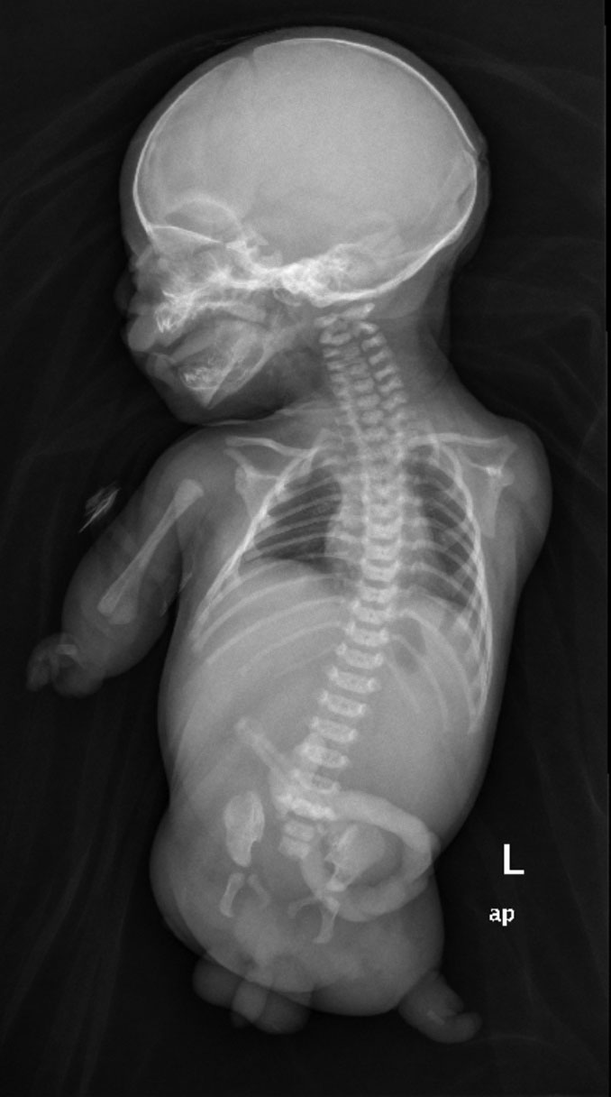
The mother was a 22 year-old healthy primigravida woman who presented for her visit at 26 weeks of gestation. The father was a healthy 23 year-old man, and there was no consanguinity or relevant family history. There was no known teratogenic exposure during pregnancy. The ultrasonographic evaluation revealed absence of both upper and lower limbs except the right humerous. Tiny stomach and bilateral mild pelvi-ureteric junction dilatation was noted, but the rest of the baby appeared normal. The couple was counseled about the poor prognosis and amniocentesis was offered but they declined. They opted for no cesarean delivery for fetal indication. Induction of labor was offered at 34 weeks of gestation, but the mother opted to wait for spontaneous labor. The mother went into spontaneous labor at 41 weeks and delivered by assisted vaginal breech delivery; a fresh stillbirth weighing 2215 grams. A baby gram was performed after birth. (Fig. 1)

Figure 1: The baby gram.

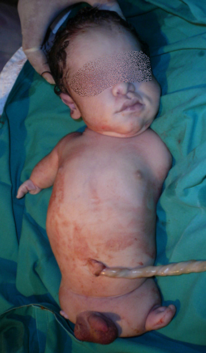
Figure 2: External examination of the stillborn baby.
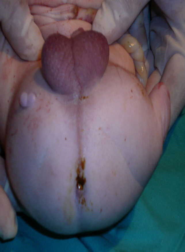
On clinical examination, there were no gross facial anomalies. Two lower limb buds and absent left upper limb was noted. The right upper limb was present up to the elbow with rudimentary palm/fingers attached to it and no forearm noted, (Fig. 2). Both testes were palpable; the anus and spine were normal. (Fig. 3)

Figure 3: Examination of the genital area of the stillborn baby
Discussion
This report presents an infant with Amelia of three limbs and/phocomelia of one limb with no striking dysmporhic features noticed. The extreme rarity of this congenital anomaly has stimulated us to report this new case presented at our hospital. Amelia was traditionally thought to be a sporadic anomaly with little risk of recurrence, or evidence of genetic origins. However, different modes of inheritance has been involved in the etiology of Amelia including autosomal recessive, X linked dominant and autosomal mode of inheritance which indicate the genetic heterogeneity of this condition.11
This report describes a case of Amelia diagnosed in a pregnancy of a non-consanguineous couple, at 26 weeks prenatal scan. Clinical examination was consistent with the prenatal findings. Amelia with multiple malformations resembling Robert syndrome have been reported earlier by song et al.12
Although Robert syndrome, which is an autosomal recessive syndrome is considered as a single genetic entity and includes various morphologic defects; babies are being reported nowadays with severe facial defects, tetra-Amelia, and pulmonary abnormality, yet with normal chromosomal findings.12 To the best of our knowledge; this is the first reported case of Amelia in the sultanate of Oman. However, Amelia, lung hypoplasia, and cleft lip palate were found to be transmitted in an autosomal recessive fashion in Arabs and Turkish families.12
In the current case; the prenatal ultrasound did not reveal gross anomalies apart from mild bilateral renal pelvis dilatation and the presence of a tiny stomach. Absent bowel gas noted on the skeletal survey after birth may have indicated a form of esophageal atresia. Infants with Amelia only appear to have good prognosis; whereas most newborns with Amelia associated with organ malformation die in the first year of life. As organ malformations have been reported to be frequently associated with cases of Amelia,12 autopsy findings would have been helpful; but unfortunately, pathological examination and chromosomal analysis were refused by the parents. The current evidence on this topic is uncertain and the exact categorization of the condition is made more difficult by the absence of autopsy.
The possibility of the recurrence of amelia has been documented in only a few families.13-15 In this case, pregnancy and family history were non-contributory factors regarding genetic or teratogenic causes; maternal infection also appears to be unlikely. It is difficult to progress further in the etiology of this case. These parents were counseled for a low recurrence rate and advised to have an early anomaly scan in future pregnancies.
Conclusions
Prenatal diagnosis including detailed ultrasound and amniocentesis play a major role in counseling parents with fetal anomalies.
Acknowledgements
The authors reported no conflict of interest and no funding was received for this work.
References
1. Lenz W. Genetics and limb deficiencies. Clin Orthop Relat Res 1980 May;(148):9-17.
2. Froster-Iskenius UG, Baird PA. Amelia: incidence and associated defects in a large population. Teratology 1990 Jan;41(1):23-31.
3. Evans JA, Vitez M, Czeizel A. Patterns of acrorenal malformation associations. Am J Med Genet 1992 Nov;44(4):413-419.
4. Mastroiacovo P, Källén B, Knudsen LB, Lancaster PA, Castilla EE, Mutchinick O, et al. Absence of limbs and gross body wall defects: an epidemiological study of related rare malformation conditions. Teratology 1992 Nov;46(5):455-464.
5. Smithells RW, Newman CG. Recognition of thalidomide defects. J Med Genet 1992 Oct;29(10):716-723.
6. Pauli RM, Feldman PF. Major limb malformations following intrauterine exposure to ethanol: two additional cases and literature review. Teratology 1986 Jun;33(3):273-280.
7. Van Allen MI, Curry C, Walden CE, Gallagher L, Patten RM. Limb-body wall complex: II. Limb and spine defects. Am J Med Genet 1987 Nov;28(3):549-565.
8. Bruyere HJ Jr, Viseskul C, Opitz JM, Langer LO Jr, Ishikawa S, Gilbert EF. A fetus with upper limb amelia, “caudal regression” and Dandy-Walker defect with an insulin-dependent diabetic mother. Eur J Pediatr 1980 Aug;134(2):139-143.
9. Bod M, Czeizel A, Lenz W. Incidence at birth of different types of limb reduction abnormalities in Hungary 1975-1977. Hum Genet 1983;65(1):27-33.
10. Källén B, Rahmani TM, Winberg J. Infants with congenital limb reduction registered in the Swedish Register of Congenital Malformations. Teratology 1984 Feb;29(1):73-85.
11. Morey MA, Higgins RR. Ectro-amelia syndrome associated with an interstitial deletion of 7q. Am J Med Genet 1990 Jan;35(1):95-99.
12. Song SY, Chi JG. Tri-amelia and phocomelia with multiple malformations resembling Roberts syndrome in a fetus: is it a variant or a new syndrome? Clin Genet 1996 Dec;50(6):502-504.
13. Zimmer EZ, Taub E, Sova Y, Divon MY, Pery M, Peretz BA. Tetra-amelia with multiple malformations in six male fetuses of one kindred. Eur J Pediatr 1985 Nov;144(4):412-41.
14. Gershoni-Baruch R, Drugan A, Bronshtein M, Zimmer EZ. Roberts syndrome or “X-linked amelia”? Am J Med Genet 1990 Dec;37(4):569-572.
15. Rodríguez JI, Palacios J, Urioste M, Rodríguez-Peralto JL. Tetra-phocomelia with multiple malformations: X-linked amelia, or Roberts syndrome, or DK-phocomelia syndrome? Am J Med Genet 1992 Jun;43(3):630-632.
|