|
INTRODUCTION
Miasis is the infestation of tissues or organs of animals or man by fly larvae. Human ophthalmomyiasis is mainly seen in people who are in contact with animals, in particular sheep and goats. The sheep nasal botfly, oestrus ovis, is by far the commonest cause of human ophthalmomyiasis. The normal hosts for the larvae are sheep and goats. Occasionally, humans are the intermediate hosts.
CASE REPORT
A 9-years-old boy presented to the Outpatient Department in February 2009 with a 1-day history of foreign body sensation and excessive watering from his right eye. He developed these symptoms after being struck in his right eye by a fly. He was absolutely fine before that and had no significant ocular or medical history. On examination; his visual acuity was 6/6 in both eyes. Examination of his left eye was normal. Eyelids of the affected right eye were normal. The conjunctiva was mildly congested with profuse lacrimation in the right eye. Five Larvae were removed with cotton buds from the conjunctiva, preserved in 70% alcohol and sent to the Veterinary Research Centre, Ministry of Agriculture and fisheries, Muscat for identification. Topical antihistaminic and antibiotic drops were given to the patient. The patient came for a follow-up after two days and he was completely relieved of his symptoms. Examination of both his eyes was normal. The larvae were identified as the first instar of Oestrus Ovis. (Fig. 1)
DISCUSSION
Ophthalmomyiasis is classified as ophthalmomyiasis externa if the larvae are present in the conjunctiva and ophthalmomyiasis interna when there is intraocular penetration of larvae.1 The symptoms of ophthalmomyiasis externa are of acute conjunctivitis with soreness, lacrimation and conjunctival congestion.
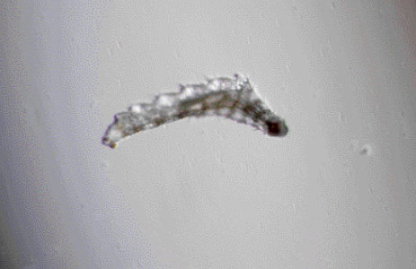
Figure 1: First instar of oestrus ovis
The symptoms of rhinitis in association with conjunctival reaction have been reported.2 External ophthalmomyiasis can be complicated by corneal ulcer, decreased vision or invasion into eye globe. Apart from mechanical removal, there is no other therapy described. Topical antibiotics are useful in preventing secondary infection by bacteria. Cases of ophthalmomyiasis externa have been reported from various parts of the world.1,3 To the best of our knowledge, this represents the third case reported from Oman of ophthalmomyiasis caused by Oestrus Ovis.4,5
CONCLUSION
Doctors in Oman should suspect ophthalmomyiasis in unilateral conjunctivitis with a foreign body sensation.
ACKNOWLEDGEMENTS
The author reported no conflict of interest and no funding was received on this work.
|