
Amna Al-Futaisi*, Faisal Al-Azri†, Anurada Ganesh**,
Khoolod Al- Mukhaini*, R.L. Koul* and Manjusha Heera*
Department of Child Health*, Department of diagnostics and molecular imaging†, and Department of Ophthalmology**. sultan Qaboos university hospital
A 13 week old female child presented with history of abnormal movements in the form of extension of the neck and stiffness of the body since five days of age. Brain Magnetic Resonance Imaging (MRI) and eye examination with fundus pictures are shown below:
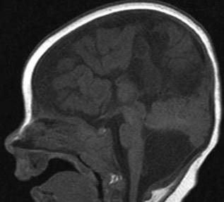
Figure 1: Sagittal T1W, midline show complete agenesis
of the corpus collasum. There is posterior fossa interhemispheric cyst.
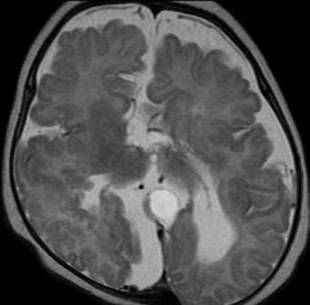
Figure 2: Corona T2W shows interhemispheric cyst, posterior fossa
arachanoid cyst and periventricular heterotopic gray matter bilaterally.
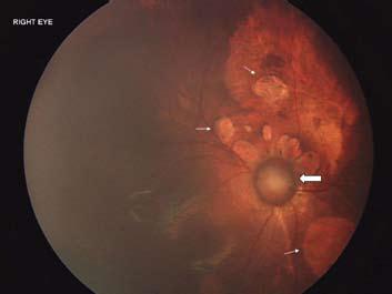
Figure 3. Small arrows show multiple well demarcated lacunae concentrated
around the optic disc. Block arrow shows optic disc coloboma.
What is the most likely diagnosis?
- Rett syndrome
- Angelman syndrome
- Tuberous sclerosis
- Aicardi syndrome
- Anderman syndrome