
The Lemon Story-An Unusual Phytobezoar causing Acute Small Bowel Obstruction
Vuthaluru Seenu,1 Ganapathy Iyer,2 Chalamalashetti S. Rao2
Seenu V, et al. OMJ. 25 (2010); doi:10.5001/omj.2010.22
ABSTRACT
Acute small intestinal obstruction is a frequent cause of admission to surgical wards. Adhesions, hernia and neoplasms are the usual causes. Phytobezoar causing obstruction is rare and acute small bowel obstruction secondary to a whole fruit is extremely rare. This is a report of acute small bowel obstruction in a 70 year old male caused by a whole undigested lemon. The diagnosis could only be established at laparotomy. Preoperative diagnosis of the exact etiology in such patients is difficult and needs a high index of suspicion. However, even when diagnosed only at laparotomy, the outcome is often successful.
From 1Department of Surgery, Oman Medical College, Sohar, Sultanate of Oman, 2Department of Surgery, Sohar Hospital, Sultanate of Oman.
Received: 12 Nov 2009
Accepted: 23 Dec 2009
Address correspondence and reprint request to: Dr. Vuthaluru Seenu, Department of Surgery, Oman Medical College, Sultanate of Oman Email: varnaseenu@hotmail.com
INTRODUCTION
Acute small intestinal obstruction is one of the commonest surgical emergencies. Adhesions, hernia and neoplasms are the usual causes. Phytobezoar causing acute small bowel obstruction is rare. Preoperative diagnosis is difficult and most cases are diagnosed at laparotomy. This is a case of acute small bowel obstruction in a 70 year old male caused by a whole undigested lemon
CASE REPORT
A 70 year old Omani male was admitted to the surgical ward with a history of colicky abdominal pain, abdominal distention, bilious vomiting and constipation of one day duration. The patient had no history of change in bowel habits, rectal bleeding or weight loss. There was no history of previous surgeries. General physical examination showed a thin, edentulous, elderly male with normal vital signs. Abdominal examination revealed mildly distended, soft abdomen with vague tenderness in all quadrants. Bowel sounds were exaggerated but rectal examination did not reveal any abnormality.
Laboratory investigations were within normal limits. Plain X-ray of the abdomen revealed multiple air and fluid levels with distended jejunal loops. Ultrasound examination of the abdomen showed dilated small bowel loops.
Considering the age and general condition of the patient, and it being the first episode of acute intestinal obstruction, a clinical diagnosis of acute intestinal obstruction secondary to a suspected malignant etiology was entertained. As the patient continued to have severe colicky abdominal pain and persistent bilious vomiting relieved only partially, by continuous nasogastric aspiration, he was taken up for emergency laparotomy, after obtaining informed consent.
Laparotomy was performed with a mid mid-line incision. At laparotomy, a 4 cm, firm, smooth surfaced swelling was palpable in mid jejunum. The bowel proximal to the swelling was dilated and the distal segment was collapsed. With gentle kneading and maneuvering, the swelling could be milked proximally. However, it could not be pushed distally into the collapsed bowel. Considering an intra luminal cause of obstruction (a pedunculated polyp/ neoplasm) or a foreign body, an enterotomy was performed, which revealed a 4 cm, whole, undigested lemon. (Fig. 1)
The entire small bowel was examined for strictures or any other foreign bodies. Following the removal of the lemon, the enterotomy was closed in two layers. Post operative recovery was uneventful and patient was discharged on the fourth post operative day.
When the lemon was shown to the patient, he denied swallowing the lemon while eating.
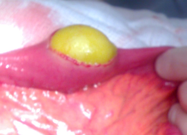
Figure 1: Shows the fresh undigested lemon protruding through the enterotomy in the jejunum. .
Discussion
Small bowel obstruction is a frequent cause of admission to surgical wards and may require emergency surgical intervention.1 The common causes of small bowel obstruction are adhesions (60%), external hernias (15%), neoplasms (6%) and inflammatory lesions (5%). Rarely, in children, elderly patients with dental prosthesis, alcoholics, prison inmates and psychiatric patients, acute small intestinal obstruction can be caused by ingested foreign bodies (FB) including food bolus (bezoars).2 Food bolus impaction is common with meat and fish bones.3,4,5 Very few cases caused by phytobezoars have been reported and food bolus obstruction due to a whole fruit is extremely uncommon. Food bolus causing acute intestinal obstruction is often seen in elderly patients with poor dentition or inadequate mastication.3 It is also more common in rice eating populations where culturally, people swallow large balls of rice mixed with large chunks of meat and vegetables. The patient in this report was also elderly and edentulous. Although, the patient denied swallowing a whole lemon, he was habituated to swallowing large balls of rice, meat and vegetables.
Majority of ingested FB pass through the gastrointestinal tract spontaneously.5 But if they get impacted in the gastrointestinal tract, they can cause obstruction, haemorrhage, perforation or fistula formation. FB can get impacted at sites of physiological narrowing in the gastrointestinal tract, constrictions in the oesophagus (arch of aorta, bronchus, and lower end), distal ileum, and ileocaecal junction or at any pathologically strictured sites. The impact of ingested FB depends on the physical characteristics such as the length and diameter of the foreign body. Spherical FB larger than 2.5 cm in diameter, are less likely to pass beyond the pylorus whereas those longer than 6 cm or even a smaller diameter, are likely to get impacted in the second or third part of the duodenum.4
The presenting symptoms of impacted bezoar vary depending upon the site of impact, type of FB and the presence or absence of complications. The common presenting symptoms are chest pain, odynophagia and respiratory symptoms (in the oesophagus), symptoms of gastric outlet obstruction (in the stomach), classical symptoms of abdominal pain, vomiting, abdominal distension and constipation (in the small bowel).
Plain X-rays of the abdomen can diagnose acute intestinal obstruction with a high sensitivity (86%).6 However, plain films cannot detect bezoars which are not radio-opaque.1 Ultrasound examination of the abdomen can occasionally identify the bezoar as an echogenic intraluminal mass. But in situations of acute intestinal obstruction, its utility is limited by the gas filled bowel loops. CT scan is helpful in diagnosing and detecting the etiology as bezoar in 73-95% of cases.3,7
Bezoars in the oesophagus and stomach can be successfully removed endoscopically. Most bezoars that have passed beyond the pylorus, pass through the rest of gastrointestinal tract and are excreted within a mean duration of four days.1 Ingested blunt FB distal to the stomach can be monitored by weekly abdominal X-ray evaluation. Sharp objects need daily radiological evaluation. Indications for surgical intervention are blunt FB remaining in the same place for more than a week, sharp object remaining in the same place for three days or the development of complications. Impacted enteral FB can be managed by open or laparoscopic approaches.1 At operation, attempt at milking the FB into the caecum can be attempted for small and blunt FB. These will be passed spontaneously in stools in 2-3 days. However, majority of the impacted bezoars need enterotomy for their removal. Tushar et al. reported a case of small bowel obstruction with an apricot managed successfully by laparoscopy.1
Discussion
In conclusion, preoperative diagnosis of bezoar as the etiology for acute small bowel obstruction is difficult and requires a high index of suspicion. The setting of elderly, demented and edentulous patients with food habits of devouring large boluses of rice, meat and vegetables should prompt the diagnosis. Management involves open or laparoscopic approaches with successful outcomes in a vast majority of cases.
References
1. Samadhi T, Signal T, Balakrishnan S, Hussain A, Grandy-Smith S, El-Hasani S. An Apricot story: view through a key hole. World J Emer Surg 2007, 2:20-21.
2. Long JWG, Austin RCT, Winslet MC: Unusual causes of small bowel obstruction .J R Soc Med 2000; 93:365-368.
3. Lerma MA, Mariscal JME, Cordon FD, Abril AG, Oron EM, Perez MJM. Small bowel obstruction caused by snail’s shell: Radiographic and CT findings. J Comp Ass Tomography 2002; 26: 529-531.
4. Eisen GM, Baron TH, Domnitz JA, Faigel DO, Goldstein JL, Johanson JF, et al. Guide lines for the management of ingested foreign bodies. Gastrointest Endosc 2002; 55:802-806.
5. Ihara N, Yashiro N, Kinoshita T, Yoshigi J, Kasai T. Small bowel obstruction due to pickled apricot: C T findings. Comp Ass Tomography 2002; 26:132-133.
7. Maglinte DDT, Balthazar EJ, Kalvin FM. The role of radiology in the diagnosis of small bowel obstruction A J R 1997; 168:1171-1180.