
Perioperative Anesthetic Management of a Case with Severe Dilated Cardiomyopathy
Mohamed A. Daabiss, Adel Hasanin
Daabiss MA, et al. OMJ. 25 (2010); doi:10.5001/omj.2010.20
ABSTRACT
Dilated cardiomyopathy (DCM) is characterized by dilatation and impaired systolic function of one or both ventricles. Five to eight people per 100,000 develop this disorder each year. It is more common in men. Malignant arrhythmias are the most common cause of death in DCM1. Around 50% of cases of nonischaemic dilated cardiomyopathy are idiopathic. Other causes are familial, infectious, infiltrative and connective tissue diseases. This is a report of successful anesthetic management of a patient with severe DCM undergoing a surgical procedure using combined thoracic epidural analgesia (TEA) and general anesthesia (GA).
From the Department of Anesthesia, Riyadh Military Hospital, Saudi Arabia
Received: 03 Oct 2009
Accepted: 05 Dec 2009
Address correspondence and reprint request to: Dr. Mohamed Daabiss, Department of Anesthesia, Riyadh Military Hospital, P.O.Box 7897- D186, Riyadh 11159, Kingdom of Saudi Arabia Email: madaabiss@yahoo.com
INTRODUCTION
Dilated cardiomyopathy (DCM) is characterized by dilatation and impaired systolic function of one or both ventricles. Each year, this disorder affects approximately five to eight people per 100,000.1It most commonly affects males than females. Malignant arrhythmias are the most common cause of death in DCM.1 Approximately 50% of cases of nonischaemic dilated cardiomyopathy are idiopathic. This is a report of successful anesthetic management of a patient with severe DCM undergoing a long surgical procedure using combined thoracic epidural analgesia (TEA) and general anesthesia (GA).
CASE REPORT
A 68 yr old male patient of 161 cm height and weighing 60 kg, who was a known case of idiopathic DCM (American Society of Anesthesia (ASA) class IV), having malignant tumor in the left kidney was scheduled for nephroureterctomy. Two weeks prior to surgery, the patient had an attack of palpitation followed by loss of consciousness which was diagnosed as congestive heart failure. 12 lead ECG and 24 hours Holters’ monitoring showed significant ventricular ectopic activities. Chest radiography showed cardiomegaly and pulmonary congestion, (Fig. 1). Echocardiography showed a severely dilated left ventricle with global hypokinesia and severe reduction of left ventricular systolic function, Ejection Fraction (EF) 20% with mitral and tricuspid regurgitation. The cardiologist treated him with carvedilol, amiodarone, captopril and spironolactone. Anticoagulant medications were not administered preoperatively.
Preoperatively, the patient was reviewed again by a cardiologist and echocardiography was repeated. The patient’s cardiac condition
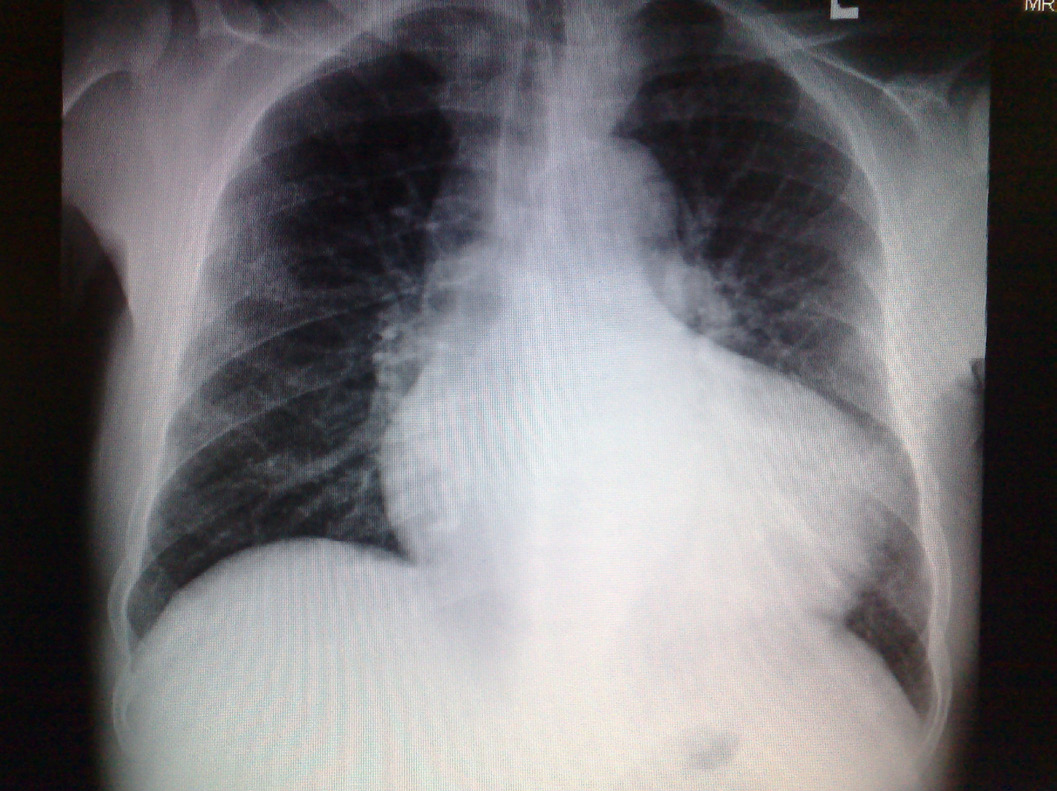
Figure 1: Chest radiography showed cardiomegaly and pulmonary congestion
condition was optimized, and the patient was not in distress or afebrile. Captopril and spirinolactone were omitted on the day of surgery. All laboratory results were within normal values. The surgical plan was to proceed into two stages at the same sitting, endoscopic laser ureteric orifice avulsion followed by laparoscopic nephrectomy. The expected operative time for both procedures was 8-9 hours. Based on the patient’s critical cardiac condition, open rather than laparoscopic nephrectomy was preferred. The patient was premedicated with lorazepam 1 mg orally 2 hours preoperative. Upon arrival into the operating theatre, the patient’s blood pressure was 120/70 mmHg, heart rate (HR) was 66/min and oxygen saturation (SaO2) was 99% on oxygen face mask 5L/min. A 14G intravenous (IV) cannula, a 20G radial artery cannula and a triple lumen catheter in the right internal jugular vein were established under local anesthetic infiltration. After positioning the patient on his right lateral side, a thoracic epidural catheter was inserted at D10-11. Bupivacaine 0.25% 10 ml with 50 μg fentanyl were titrated over 2-3 minutes through the epidural catheter. The patient was kept supine, head and shoulder up 30° with O2 face mask 5L/min. Sensory loss was at T10 dermatome. Dopamine infusion was started of 3-5μ/kg/min through the central venous line. Intraoperatively, the mean arterial blood pressure (MAP) was 85-105mmHg, HR was 57-66 beats/min, central venous pressure (CVP) was 10-17 cmH2O and SaO2 was 99-100%. Sedation was achieved with IV midazolam 1mg. An epidural top up of 5 ml bupivacaine 0.25% with 25μg fentanyl was titrated to maintain the level of sensory loss. Several arterial blood samples were analyzed for blood gases (ABG) and electrolytes, which were within normal values. Total fluids received were 1200 ml of crystalloids in addition to about 500 ml of absorbed irrigation crystalloid fluid used in 4 hours endoscopic ureteric procedure. Preparing for the second stage of surgery, 6 ml bupivacaine 0.5% added to 50 μg of fentanyl were titrated through the epidural catheter to reach analgesic level to T8. After that, general anesthesia (GA) was induced using IV injection of etomidate 10 mg and fentanyl 50 μg. Vecronium bromide 6 mg was given to facilitate the insertion of a cuffed endotracheal tube. Anesthesia was maintained with isoflurane (0.5-1 MAC) O2/Air and intermittent vecuronium bromide. Dopamine infusion was used to maintain adequate MAP. The patient was positioned in lateral loin position for nephrectomy. This stage lasted 3 hours during which MAP was 80-95 mmHg, HR was 60-71 beats/min, SaO2 was 99-100%, CVP was 9-15 cmH2O, end tidal carbon dioxide was 33-37 mmHg and airway pressure was 15-18 cmH2O. ABG and electrolytes were within normal levels. ECG tracing was normal sinus rhythm with infrequent PVCs (<6 /min) without any ischemic changes. Total urine output was 500 ml. At the end of the surgery, the residual muscle relaxant effect was reversed and patient was extubated smoothly. Overall, the anesthetic management was uneventful. In the PACU, the patient was fully awake and on epidural infusion of bupivacaine 0.03% with fentanyl 4 μg/ml, 3-5 ml/hour for postoperative pain control. After an hour, the patient was transferred to High Dependency Care Unit with stable vital signs. The following day, the patient was discharged to the surgical ward with stable hemodynamics.
DISCUSSION
The present case had two major problems, DCM with severe cardiac dysfunction (EF 20%) and prolonged surgical procedure (7 hours) with the associated hemodynamic and intravascular volume changes. The goals for anesthetic management were avoidance of drug induced myocardial depression, maintenance of normovolemia, and prevention of increased ventricular afterload. GA solely may increase the risk of CHF, myocardial ischemia or intraoperative arrhythmias.3 El-Dawlatly et al. reported uneventful anesthetic management of a patient with DCM who underwent laparoscopic cholecystectomy (LC) under TEA.3 Aono et al. compared three anesthetic techniques: GA, epidural analgesia (EA) and GA combined with EA for LC. They reported that, GA with sevoflurane/N2O could not suppress stress response of both hypothalamus-pituitary-adrenocortical axis and sympathoadrenal system while EA suppressed only the symathoadrenal responses.4 They concluded that TEA may be of advantage in patients with limited cardiac function undergoing abdominal surgery. Gramatica et al. used EA as a sole technique for LC and recommended it for patients who are not good candidates for GA due to cardiorespiratory problems.5 The choice for anesthetic management in this case was fentanyl based EA with low concentration local anesthetic agent. This technique offers satisfactory analgesia with a relatively slow sympathetic blockade as well as decreased peripheral vascular resistance. In addition, induced reduction in afterload and preload benefits cardiac function.6 Hashimoto et al. reported that high dose epidural fentanyl anesthesia is an anesthetic method of choice for patients with DCM.7 The small dose of dopamine used in this case was to support the circulation if hypotension occurred in light of the cardiac compromised status of the patient. Changing the surgical plan to open laparotomy reduced the risk of pnuemoperitonieum e.g. hypercarbia and cardio-pulmonary compromise.8 In the second stage of surgery, the patient was positioned in lateral loin position with the added risk of pleural injury during dissection due to tumor infiltration. Therefore, EA would not be satisfactory unless combined with relatively light GA to offer good analgesia with hemodynamic stability. Monitoring of CVP with the other vital signs was used to optimize the preload. CVP was kept between 12-16 cmH2O and MAP 80-95mmHg. Although cardiac output was not measured intraoperatively, it was believed that the circulatory effects of epidural block contributed to a relatively stable operative and postoperative course. Swan Ganz catheter was not used as the patient position (lateral loin) might have disrupted the tracing of pulmonary capillary wedge pressure (PCWP) as well as cardiac output figures, and therefore eliminates the advantage of its use in addition to its coexisting complications. This is in agreement with Amaranath et al. and Kanu.6,9 Although using Transesophageal Echocardiography (TEE) could be useful, it was not used due to limited expertise with TEE. To the best of our knowledge, this may have been one of the longest cases reported on the use of TEA with GA in patient with severe cardiomyopathy. Such patients could be well managed with preoperative optimized medical condition and well-planned anesthetic management.
CONCLUSION
In conclusion, TEA with relatively light GA offers good analgesia with hemodynamic stability for major abdominal surgery in patients with critical cardiac condition.
ACKNOWLEDGEMENTS
The authors reported no conflict of interest and no funding was received on this work.
REFERENCES
1 Dec GW, Fuster V. Idiopathic Dilated Cardiomyopathy. N Engl J Med, 1994; 331:1564-1575.
2. Stoelting RK, Dierdorf SF. Cardiomyopathy, In: Stoelting RK (ed). Anesthesia and Coexisting Disease (3rd ed). New York: Churchill Livingstone 1993; 97-102.
3. El-Dawlatly A, Al-Dohayan A, Fadin A. Epidural Anesthesia For Laparoscopic Cholecystectomy In A Patient With Dilated Cardiomyopathy: Case Report And Review Of Literature. Internet J Anesthesiol. 2007; 13(1).
4. Aono H, Takeda A, Tarver SD, Goto H. Stress responses in three different anesthetic techniques for carbon dioxide laparoscopic cholecystectomy. J Clin Anesth 1998; 10:546-550.
5. Gramatica L, Brasesco OE, Mercado LA, Martinessi V, Panebianco G, Labaque F, et al. Laparoscopic cholecystectomy performed under regional anesthesia in patients with chronic obstructive pulmonary disease. Surg Endosc 2002; 16:472-475.
6. Amaranath L, Shahpour E, Lockrem J, Rollins M. Epidural analgesia for total hip replacement in a patient with dilated cardiomyopathy. Can Anaesth Soc J 1986; 33(1):84-88.
7. Hashimoto K, Ooka T, Kosaka Y. Epidural anesthesia with high dose fentanyl for a patient with dilated cardiomyopathy. Masui. 1994 ; 43(12):1881-1884.
8. Jansen F W, Kapiteyn K, Trimbos-Kemper T, Hermans, J Trimbos, JB. Complications of laparoscopy: a prospective multicentre observational study. Br J Obstet Gynecol,1997; 104:595-600.
9. Kanu C. The Swan-Ganz Catheters: Past, Present, and Future, A Viewpoint. Circulation, 2009; 119:147-152.