
Encysted Hydrocele of Cord in an Adult Misdiagnosed as
Irreducible Hernia: A Case Report
Imtiaz Wani, Muddasir Rather, Gulam Naikoo, Imran Gul, Zubair Bhat, Aejaz Baba
Wani I, et al. OMJ. 24, 218-219 (2009); doi:10.5001/omj.2009.42
ABSTRACT
A number of pathologies can present as groin swellings in adults.Among these, encysted hydrocele of the cord presenting as swelling in an adult is a rare. A case of encysted hydrocele of cord in 36 year old male mimicking as as an irreducible hernia is reported. The diagnosis of hydrocele was made intraoperatively. An excision of the sac was performed.
Department of surgery ,S.M.H.S Hospital /SKIMS Srinagar
Received: 27 Feb 2009
Accepted: 16 Mar 2009
Author for correspondence: Imtiaz Wani, Amira Kadal, Srinagar, Kashmir, India.
e-mail:imtazwani@gmail.com
CASE REPORT
A 36 year old male was presented with swelling in the right groin for a 12 days duration. For the last 2 days, the patient had severe progressive pain in groin area and low grade fever for which he reported to the emergency services. There was no history of constipation, loose motions or vomiting. General physical examination as well as systemic examination were normal. Abdominal examination was normal, with no organomegaly and normal bowel sounds were present.
On local examination, a globular, soft, tender swelling measuring 5×2.3×1 centimeter, with negative cough impulse was present in the right inguinal region (Fig. 1).
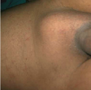
Figure 1: Swell in Right Groin
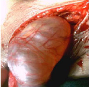
Figure 2: Sac in Right Groin
The swelling could be felt completely separate from the testicle. Transillumination test as well as traction test was negative. Genitilia examination was normal. Per rectal examination was unremerkable. All baseline investigations were normal with a hemoglobin level of 13 gm/dl., total leucocyte count of 7,500/ mm3, and normal electrolytes. An x-ray of the abdomen did not reveal any evidence of intestinal obstruction. Ultrasongraphy of the abdomen was normal. Scrotal ultrasound showed an oval anechoic mass in the groin. The patient was managed by intravenous fluids, antibiotics and pain killers but had mild relief of symptoms and diagnosis of irreducible hernia was made. The patient had exploration of the right groin and a sac was found abutting the spermatic cord having flimsy adhesions with the surrounding tissues (Fig. 2). The sac was abutting spermatic cord at the proximal end of the sac starting about 2 centimeters from the deep inguinal ring with no scrotal extension observed. Aspiration of the contents of the sac revealed an amber colored fluid. An excision of the sac was performed. Fluid analysis was consistent with that of hydrocele fluid. Histopathological examination of the cyst wall showed collagenous material. Postoperative period was uneventful and the patient is regularly attending follow up clinics visits.
DISCUSSION
The main pathological conditions manifesting as masses in the groin fall into five major groups: congenital abnormalities, non-congenital hernias, vascular conditions, infectious or inflammatory processes, and neoplasms.1 Inflammatory swellings of the groin are common, and the changes are often attributed to infection and are often inflammatory swellings secondary to groin hernia.2 However, painful spermatic encysted hydrocele presenting as a groin swelling is rare.
An encysted hydrocele or a non-communicating type of inguinal hydrocoele, is a loculated fluid collection along the spermatic cord, separated from and located above the testicle and the epididymis, as a result of aberrant closure of the processus vaginalis. This is idiopathic in most cases but in some cases it may be secondary to testicular torsion, tumour or trauma, and in infections, as in, orchitis, epididymitis, tuberculosis or filariasis.3 Rarely, hydrocele of pancreatic origin have been reported to occur.4 Encysted hydrocele of the cord remains asymptomatic or is detected incidentally during evaluation during the course of other disease.5
Diagnosis is clinicaly essential but where doubt exists, scrotal ultrasound can be used to differentiate it from other scrotal lesions. Diagnosis can also be confirmed by computed tomography scan or intraoperatively. Spermatic cord hydrocele is effectively diagnosed by ultrasonography based on its specific location and shape. Ultrasonography is useful to exclude hernia, enlargement of the lymph node, or other solid masses.6 A typical finding on ultrasonography of spermatic cord hydrocele is its avascular anoechoic structure.
Excision is the treatment of choice and the excision under local anesthesia in adult patiens is well studied.7 Fluid analysis of the hydrocele fluid showed amber color and sterile in nature Specific gravity of the fluid was 1.02. Microscopically, cholesterol crystals were isolated with tests positive for presence of albumin and fibrinogen. Histopathological examination of the cyst wall shows collagenous material. Encysted type can be misdiagnosed as hernia, lymphagiomatous cyst or cystic teratoma, inguinal lymphadenopathy, lipoma of cord ,or other tumours of the cord. Rarely, ileo femoral aneurysm, appendicular pathology, or a hematoma present as an inflammatory swelling in the groin.8
CONCLUSION
Encysted hydrocele of the cord in an adult is a rare condition .It may mimic an irreducible hernia at times. Excision remains the treatment of choice.
ACKNOWLEDGEMENTS
The authors reported no conflict of interest and no funding has been received on this work.
-
Shadbolt CL, Heinze SBJ, Dietrich RB. Imaging of Groin Masses: inguinal anatomy and pathologic conditions revisited. RadioGraphics. 2001;21:s261–71.
-
Maheswaran P, Stephen D. A rare presentation of appendicitis as groin swelling: a case report. Cases J. 2009; 2: 53.
-
Ku HJ, Kim ME, Lee NK, Park YH. The excisional, placation and internal draingetechniques: a comparison of the results for idiopathic hydrocele. BJU Int. 2001;87:82–84.
-
Delamarre J, Descombes P, Grillot G, Deschepper B, Deramond H. Hydrocele of pancreatic origin. X-ray computed tomographic study of an intrascrotal collection in an acute outbreak of chronic pancreatitis. J Radiol. 1988;69:689-90.
-
Busigo J. P, Eftekharif F, Hendell H .Encysted Spermatic Cord Hydroceles : A Report of Three Cases in Adults and a Review of the Literature. Acta Radiologica. 2007; 48:1138-1142.
-
Han BH, Cho JY, Cho BJ, Ki WW Hydrocele of the Spermatic Cord: Ultrasonograhic Findings. J Korean Soc Med Ultrasound. 2002 ;21:129-133.
-
Agbakwuru A , Salako A, Olajide A, Takure AO, Eziyi A. Hydrocelectomy under local anaesthesia in a Nigerian adult population Afr Health Sci. 2008; 8: 160–162.
-
Apostolidis S, Papavramidis S, Michalopoulos A, Papadopoulos N, Paramythiotis D, Harlaftis N. Groin Swelling, the Anatomic Way Out of Abdominal Haematomas Acta Chir Belg, 2008, 108, 251-253.