
Metastatic Squamous Cell Carcinoma of Foot: Case Report
Imtiaz Wani
ABSTRACT
Squamous cell carcinoma of the foot is rare. A case report of occurrence of metastatic squamous carcinoma of the foot with resemblance to verrucous type in an elderly female is presented. This metastatic lesion developed four years after excision of primary squamous cell carcinoma from the left thigh. The patient presented with a painful, exophytic, ulcerated mass on the foot of four months’ duration. Fine-needle aspiration and edge biopsy documented this lesion as squamous cell carcinoma.
From the Postgraduate Department of Surgery, S.M.H.S Hospital Srinagar.
Received: 26 Aug 2008
Accepted: 09 Nov 2008
Address correspondence and reprint requests to: Dr. Imtiaz Wani, Post graduate Department of Surgery, S.M.H.S Hospital Srinagar, India 190009.
E-mail: imtazwani@gmail.comINTRODUCTION
Squamous cell carcinoma (SCC) is relatively rare in the foot.1 This carcinoma of the foot may arise from a precursor lesion or may be secondary. Presentation can be either as a proliferative or an erosive lesion. Although most cases are curable, tumors may recur or metastasize. Squamous cell carcinoma of the foot may resemble verrucous carcinoma or there can be distinct verrucous carcinoma of the foot or epithelioma cuniculatum, usually occurring on the inner aspect of the foot, a slow-growing variant of squamous cell carcinoma with a low metastatic potential. Magnetic resonance imaging is adjuvant in the diagnosis of squamous carcinoma of the foot. Histopathology is usually diagnostic. The degree of differentiation of squamous cell carcinoma, as well as size and depth of tumor invasion are extremely important prognostic variables.
CASE REPORT
A 70-year-old female, postmenopausal, hypertensive and nondiabetic, presented to our Outpatient Department (OPD) services with a painful, ulcerated swelling on the left foot of four months’ duration. No history of trauma, infected lesion or any chronic dermatological infliction of the foot was present. The only significant past history was that the patient had had excision of a 6 cm ulcerated swelling from the right lateral thigh four years ago and had diagnosis of well-differentiated squamous cell carcinoma of the thigh. On general physical examination, pallor and inguinal lymphadenopathy were present. Systemic examination was normal. Local examination of the swelling revealed an ulcerated lesion with dimensions of 10.7×11.8×3.6 cm, tender and mobile, free from underlying tissues, extending to the sole inferiorly and to the ankle superiorly. (Figure 1)
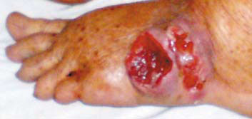
Figure 1: Foot carcinoma - pallor and inguinal lymphadenopathy
Proximal joint movement was normal. Foot X-ray showed soft tissue swellings and no apparent bone involvement. Abdominopelvic and chest computed tomography scan was normal. Multiple lymph nodes of <2 cm were palpable in the groin area and fine-needle aspiration of these lymph nodes was documented as reactive hyperplasia. Fine-needle Aspiration (FNA) and edge biopsy of swelling was consistent with squamous cell carcinoma and inguinal lymph nodes showing reactive hyperplasia. The patient refused amputation of the foot and did not return for followup.
DISCUSSION
Approximately 80% of non-melanoma skin cancers are basal cell carcinoma and 20% are squamous cell carcinoma. This squamous cell carcinoma originates from the squamous cell epithelium of surface dermis and may show varying degrees of differentiation and keratinization. In the foot, this cancer may arise from lichen planus, deep mycosis, lichen simplex chronicus, plantar verruca or this can be secondary.2 Metastatic squamous cell carcinoma of the foot is very rare. Clinical appearance of squamous cell carcinoma is variable and the tumor may present as a thin, red or brown nodule with or without scaling, a focus of induration, an ulcerated lesion plaque or an exophytic, cauliflower-like growth. A high index of suspicion is necessary to make the early diagnosis of malignancy and prevent spread of lesions; overlooking this can be disastrous, and can lead to malignancy.3 Among radiological investigations, Magnetic Resonance Imaging (MRI) helps to determine the presence and extent of disease in the skin, surrounding tissue and subjacent bone and therefore aids in surgical planning.
An initial wide excision for squamous cell carcinoma of the foot is the treatment of choice and may prevent metastasis. Inadequate excision associated with recurrence should be treated by amputation.4 Altay M et al. suggested that the treatment of choice for squamous cell carcinoma of the foot is amputation and routine lymphadenectomy at the time of management is unnecessary, but regional lymphadenectomy persisting three months after amputation warrants surgical intervention.5 The treatment that offers the highest rate of cure for patients with high-risk primary or recurrent squamous-cell carcinoma is Mohs micrographic surgery. Appropriate use of electrodesiccation and curettage, excision, or cryosurgery can eliminate up to 90% of local tumors with low risk of metastasis, especially those being less than 1 cm and are relatively inexpensive to perform. Left external iliac catheterization and intra-arterial infusion with methotrexate with salvage by leucovorin is a simple and effective method for squamous cell carcinoma of the foot with the unique advantage of preservation of organ and function.1 The five-year rate of cure in patients with large tumors is 70%, regardless of the treatment chosen.
Differential diagnosis includes keratoacanthoma, basal cell carcinoma, deep mycosis, eccrine poroma, sweat gland carcinoma, amelanotic melanoma, pyogenic granuloma, reactive epidermal hyperplasia, overlying site of infection, chronic mechanical trauma changes and cutaneous Hodgkin’s disease. In treating recalcitrant ulcers that have not responded to conventional modes of therapy, malignancy should be ruled out and biopsy done.
Metastatic potential of squamous cell carcinoma is often underestimated. Sentinel Lymph Node Biopsy (SLNB) accurately diagnoses subclinical lymph node metastasis with few false-
negative results and low morbidity. Patients who develop one squamous cell carcinoma have a 40% risk of developing additional squamous cell carcinoma within the next two years. This risk is likely even greater as more time elapses. The 90% of metastatic SCC occur within three years of diagnosis of the primary tumor. The majority of the metastatic lesions originate from primary tumors stratified in the “high-risk” category; the characteristics of high-risk squamous cell carcinoma on extremities being size >2.0 cm, indistinct clinical borders, rapid growth, multiple lesions, ulceration, recurrence after previous treatment, with histopathological documentation, poor differentiation, deep extension of the tumor into subcutaneous fat, perineural/perivascular or intravascular invasion. Metastasis may also be related to anaplastic transformation by radiotherapy. A tumor size larger than 2 cm doubles the recurrence rate and triples the metastatic rate as compared with lesions less than 2 cm. While squamous cell carcinoma developing from precursor lesions such as actinic keratoses are considered less likely to metastasize, secondary squamous cell carcinomas, which have the poorest prognosis, include tumors that develop in burn scars, in sites of radiation damage and in sites of chronic inflammation such as osteomyelitic foci and, at times, chronic leg ulcers.6
Chemoprevention with systemic retinoids is effective for reducing the number of new squamous cell carcinomas in both immunocompetent and immunosuppressed patients. Prophylactic use of oral Acitretin for treatment must be continued indefinitely because a relapse in tumor development occurs following discontinuation of oral retinoids.
CONCLUSION
Squamous cell carcinoma of foot is rare to see. It can be a primary or metastic lesion. This lesion of foot has a potential of recurrence. Wide local excision is treatment of choice.
-
Spinosa FA. Squamous cell carcinoma of the plantar aspect of the foot. J Foot Surg. 1987; 26:253-255.
-
Shahid Majeed, Bari AU. Squamous cell carcinoma foot arising in deep mycosis. A case report. J Surg Pak 2004; 9:54-55.
-
Sheen YS, Sheen MC, Sheu HM, Yang SF, Wang YW. Squamous cell carcinoma of the big toe successfully treated by intra-arterial infusion with methotrexate. Dermatologic Surgery 2003; 29:982-983.
-
Schroven I, Hulse G, Seligson D. Squamous cell carcinoma of the foot:
Two case reports. Clinical orthopaedics and related research 1996; 328:227-230. -
Altay M, Arikan M, Yildiz Y, Saglik Y. Squamous cell carcinoma arising chronic osteomyelitis in foot and ankle. Foot Ankle Int. 2004; 25:805-809.
-
Baldursson B, Sigurgeirsson B, Lindelöf B. Leg ulcers and squamous cell carcinoma. An epidemiological study and a review of the literature. Acta Derm Venereol 1993; 73:171-174.