
Unusual Skin Metastasis from Squamous Cell Carcinoma of
the Oesophagus
Gaurav Maheshwari, Nikhil Kale, Premashish Halder
Maheshwari G, et al. OMJ. 25, 51-52 (2010); doi:10.5001/omj.2010.14
From the Department of Surgical Gastroenterology
Received: 09 Dec 2009
Accepted: 05 Jan 2010
Address correspondence and reprint requests to: Dr. Gaurav Maheshwari, Jagjivanram Railways Hospital, Mumbai, India
E-mail: drgauravmaheshwari@gmail.com
Cutaneous metastases from internal malignancies are very common. This report describes an extremely rare case of cutaneous metastases from esophageal carcinoma.
This is a case of an uncommon cutaneous metastases from squamous cell carcinoma (SCC) of the esophagus in a 62-year-old female. This report emphasizes the importance that newly appearing skin lesions may be the first presentation of metastasis from esophageal carcinoma.
A 62 yr old female admitted with h/o dysphagia to solids since three months. Upper GI endoscopy showed polypoid growth in mid esophagus at 30 cm beyond which scope could not be negotiated. Biopsy was taken and reported as moderately differentiated squamous cell carcinoma.
After confirming operability and resectability transhiatal esophagectomy was performed in March 2008. Histopathological report showed moderately differentiated squamous cell carcinoma, extending up to the muscularis layer with lymphovascular metastasis, and nine out of thirteen nodes showed evidence of metastasis, however resection margins were free.
Postoperative period was uneventful with no evidence of leak and the patient was able to eat solids. In view of R0 resection and HPE diagnosis of squamous cell carcinoma, the patient was not subjected to any form of adjuvant treatment.
Patient followed up in August 2008 when she complained of a swelling on the left side of the chest wall.
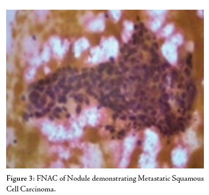
On examination, a 2 cm x 1 cm hard, nontender nodule, with no increase in local temperature, fixed to the skin but not to chest wall or ribs was palpated. There was no evidence of ulceration or discharge from the nodule. FNAC of the nodule was reported as metastatic squamous cell carcinoma.
In September 2008, PET CT scan revealed active mediastinal, abdominal and retroperitoneal nodal disease. Bilateral pleural disease and pulmonary nodules in the right lung were also present.
The patient was started on palliative chemotherapy with Taxol and Carboplatin. Four cycles were completed in December 2008. As there was no response to this treatment, the patient was then started on chemotherapy with Capecitabine plus Oxaliplatin, which again did not show any response. She was counseled and discharged on supportive treatment.
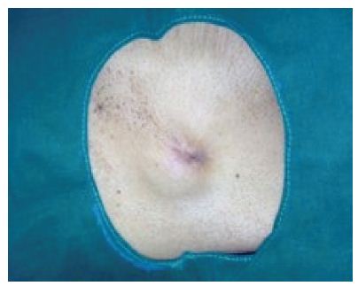
Figure 1: Site of the Metastatic Tumor Nodule on the Chest Wall.
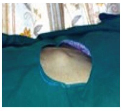
Figure 2: Another View of Nodule on Chest Wall
In general, skin metastases from malignant tumors of the internal organs are rarely seen, with a frequency of between 0.7 and 9%.1 Esophageal carcinoma rarely present with clinical features of skin metastasis. There is limited review in the recent literature
because of this rare presentation. A study by Lookingbill et al. of 7316 cancer patients with metastases to the skin had no patients with primary esophagus carcinoma.2 Another study of 4020 cancer patients by the same author found only 3 patients with cutaneous metastases from esophageal cancer, spreading mainly to the chest and abdomen as in the current case. Contrary to more common adenocarcinoma, the primary tumor in all 3 cases was of squamous cell origin.3 Fereidooni et al. also reported esophageal adenocarcinoma with facial skin metastasis.4 One additional case has been published discussing metastases on a digit from a basaloid variant of esophageal squamous cell carcinoma.5 An interesting review by Schoenlaub et al. of the clinical findings and overall survival of 200 patients with cutaneous metastases of various cancers revealed poor prognosis associated with esophageal adenocarcinoma.6 Tharakaram described five cases of skin metastases from ESCC in male patients.7
However, as newer combined-modality treatments, such as neoadjuvant chemoradiation followed by surgical resection, and newer cytotoxic agents became available, loco-regional disease recurrence has been dramatically reduced. Today, in most patients who have undergone combined-modality treatments for advanced esophageal carcinoma, recurrences have mainly been from hematogeneous metastatic spread, as in the current case. It is postulated that a subpopulation of slowly growing chemotherapy-refractory cells remained viable after the treatment; these malignant cells appear to have been deposited in the dermal lymphatic channels and remained quiescent for an extended period of time until their gross clinical presentation as skin nodules. A complete history, careful physical examination, and adjunctive radiographic studies (CT scans, bone scans, positron-emission tomography) are essential in the assessment of distant failures.
In conclusion, there is a trivial shift in pattern of metastasis from local recurrence to more distant metastasis in esophageal carcinoma. This can be explained by more stringent local attack in form of adjuvant and neoadjuvant chemoradiation. Patients may present with distant metastasis much earlier than they develop local recurrence in form of dysphagia. A complete history, careful physical examination, and adjunctive radiographic studies (CT scans, bone scans, positron-emission tomography) are essential in assessment of distant failures which suggests poor prognosis.
ACKNOWLEDGEMENTS
The authors reported no conflict of interest and no funding has been received on this work.
-
Spencer PS, Helm TN. Skin metastases in cancer patients. Cutis 1987 Feb; 39(2):119-121.
-
Lookingbill DP, Spangler N, Sexton FM. Skin involvement as the presenting sign of internal carcinoma. A retrospective study of 7316 cancer patients. J Am Acad Dermatol 1990; 22:19-26.
-
Lookingbill DP, Spangler N, Helm KF. Cutaneous metastases in patients with metastatic carcinoma: A retrospective study of 4020 patients. J Am Acad Dermatol 1993 Aug; 29(2):228-236.
-
Fereidooni F, Kovacs K, Azizi MR, Nikoo M. Skin metastasis from an occult esophageal adenocarcinoma. Can J Gastroenterol 2005 Nov; 19(11):673-676.
-
Houston JD, Telepak RJ. An isolated digital metastasis of esophageal basaloid squamous cell carcinoma. Clin Nucl Med 2000 Jul; 25(7):557-558.
-
Schoenlaub P, Sarraux A, Grosshans E, Heid E, Cribier B. Survival after the occurrence of cutaneous metastasis: a study of 200 cases. Ann Dermatol Venereol 2001 Dec; 128(12):1310-1315.
-
Tharakaram S. Metastases to the skin. Int J Dermatol 1988 May; 27(4):240-242.